
 By Suherman Candra Maholtra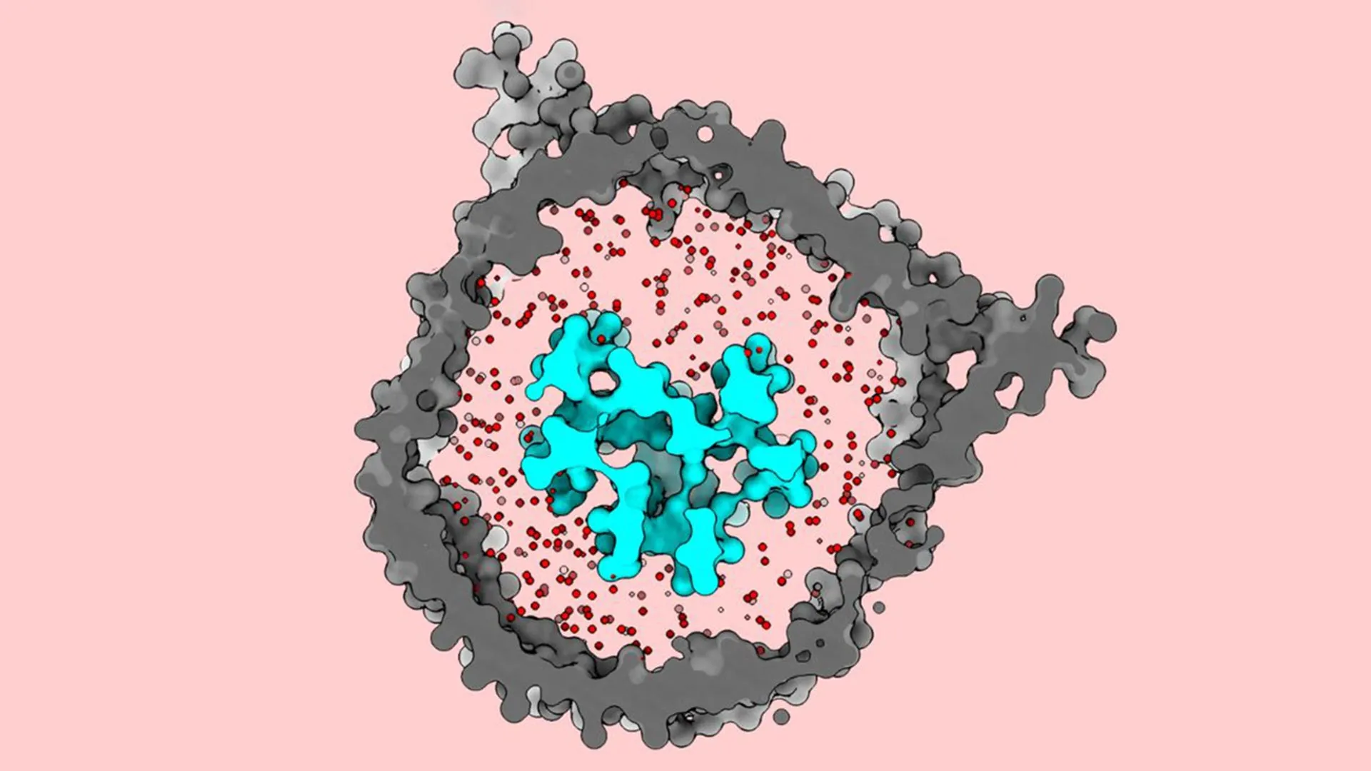
In a landmark study published in Nature Communications, scientists at The Rockefeller University have resolved a long-standing mystery regarding the structural mechanics of the T cell receptor (TCR), a discovery that could fundamentally alter the landscape of oncology and immunotherapy. By utilizing cutting-edge cryogenic electron microscopy (cryo-EM) and a novel biochemical environment that mimics the human cell membrane, the research team revealed that the TCR functions through a "jack-in-the-box" mechanism—remaining tightly coiled and dormant until it encounters a specific threat. This finding corrects decades of misconceptions based on previous structural models and provides a blueprint for engineering more effective treatments for cancers that have traditionally resisted immune-based therapies.
The Evolution and Limitations of T Cell Immunotherapy
The field of oncology has been transformed over the last decade by the advent of T cell immunotherapies. These treatments, which include Chimeric Antigen Receptor (CAR) T-cell therapy and TCR-engineered therapies, leverage the body’s natural defense system to identify and eliminate malignant cells. The underlying principle involves extracting a patient’s T cells, genetically modifying them to recognize specific cancer markers, and reintroducing them into the bloodstream.
Despite the clinical success of these therapies in treating certain blood cancers, such as B-cell acute lymphoblastic leukemia and non-Hodgkin lymphoma, their efficacy in solid tumors—including sarcomas, carcinomas, and glioblastomas—has remained stubbornly low. Scientists have long suspected that the barrier to success lies in the molecular signaling of the T cell receptor itself. However, because the TCR is a highly complex protein assembly embedded within the oily, fluid environment of the cell membrane, capturing high-resolution images of its active and inactive states has proven exceptionally difficult. Without a clear understanding of how the TCR "switches on," researchers have been forced to design immunotherapies with an incomplete map of the cellular machinery they are attempting to manipulate.
A Breakthrough in Molecular Visualization
The research was conducted at Rockefeller’s Laboratory of Molecular Electron Microscopy, led by Thomas Walz, a world-renowned expert in cryo-EM imaging. The study’s first author, Ryan Notti, brought a unique perspective to the project as both a structural microbiologist and a physician-scientist at Memorial Sloan Kettering Cancer Center. Notti’s clinical work involves treating patients with sarcomas—aggressive cancers of the bone and soft tissue that rarely respond to current immunotherapies.
To observe the TCR in its natural state, the team moved away from traditional biochemical methods. Historically, researchers used detergents to extract proteins from cell membranes for study. While effective for many proteins, detergents often strip away the surrounding lipids that provide structural support to membrane-bound complexes. Previous cryo-EM studies conducted in detergent-based environments depicted the TCR as being in a permanently "open" or extended state, leading to the conclusion that no significant structural change occurred during activation.
Walz’s lab utilized "nanodiscs"—microscopic, disc-shaped sections of membrane held together by scaffold proteins—to recreate the TCR’s native milieu. By carefully selecting a lipid mixture that mirrored the exact composition of a human T cell membrane, including specific concentrations of cholesterol and phospholipids, the researchers were able to stabilize the receptor in a way that preserved its natural behavior.
The Jack-in-the-Box Mechanism Explained
When the TCR was imaged within this restored membrane environment, the results were startling. Contrary to previous models, the dormant T cell receptor appeared compact and folded inward. It was only when the receptor was exposed to an antigen—a protein fragment presented by the Human Leukocyte Antigen (HLA) complex on a target cell—that it underwent a dramatic conformational shift.
The receptor essentially "sprang" open, extending outward from the cell surface. This structural transition is what triggers the internal signaling cascade that tells the T cell to attack. The researchers likened this to a jack-in-the-box: the "lid" is held shut by the physical constraints of the cell membrane and the specific arrangement of the TCR’s eight constituent proteins. When the correct antigen "cranks the handle," the tension is released, and the receptor unfolds to initiate the immune response.
This discovery explains why previous attempts to understand TCR signaling were flawed. Without the lipid membrane to hold the receptor in its "closed" position, the complex would naturally drift into an open state, leading scientists to believe that the extended form was its default configuration.
Chronology of the Research and Technical Challenges
The journey to this discovery began several years ago when Notti, during his doctoral studies, identified a gap between structural biology and clinical outcomes in oncology. He noted that while clinicians were attempting to "tune" T cells to be more sensitive to cancer, they lacked the structural data to know which parts of the receptor to modify.
The experimental phase of the study involved several years of meticulous protein engineering. The T cell receptor is not a single protein but an octameric complex—a cluster of eight different protein chains (alpha, beta, and the CD3 subunits gamma, delta, epsilon, and zeta) that must assemble with perfect precision.
- Protein Synthesis: The team had to produce all eight proteins and ensure they folded correctly.
- Nanodisc Integration: The proteins were then inserted into nanodiscs. This step required fine-tuning the lipid-to-protein ratio to ensure only one TCR complex was embedded per disc.
- Cryo-EM Preparation: The samples were flash-frozen in liquid ethane to preserve their structure in a "vitreous" (glass-like) ice state, preventing the formation of ice crystals that would distort the image.
- Data Collection and Analysis: Thousands of two-dimensional electron microscopy images were processed using computational algorithms to reconstruct a high-resolution three-dimensional model of the receptor.
Implications for Next-Generation Cancer Treatment
The ability to see the TCR in its dormant and active states provides a new set of targets for drug development and genetic engineering. For patients with sarcomas and other solid tumors, the problem is often that the T cells are either "blind" to the cancer or the activation threshold of the TCR is too high to overcome the immunosuppressive environment of the tumor.
"Re-engineering the next generation of immunotherapies tops the charts in terms of unmet clinical needs," Notti stated. By understanding the "jack-in-the-box" mechanism, scientists can now explore ways to adjust the "spring tension" of the receptor. This could involve:
- Sensitivity Tuning: Modifying the TCR to spring open more easily when it encounters low-density antigens on the surface of tumor cells.
- Precision Engineering: Designing synthetic receptors that remain tightly closed in the presence of healthy cells but respond with maximum force when a cancer-specific antigen is detected, thereby reducing the "off-target" toxicity that often plagues current treatments.
- Vaccine Optimization: The structural data provides a clearer picture of how different antigens interact with the TCR. This information is vital for the development of cancer vaccines, which aim to train a patient’s immune system to recognize mutations before a tumor can take hold.
Broader Impact on Immunology and Vaccine Design
Beyond cancer, the findings have significant implications for the treatment of autoimmune diseases and infectious pathogens. In autoimmune conditions, T cells mistakenly attack the body’s own tissues. Understanding the mechanics of TCR activation could allow researchers to design "molecular brakes" that keep the receptor in its closed, dormant state, preventing unwanted immune responses.
Thomas Walz emphasized that this work represents the pinnacle of basic science’s role in medicine. "The T cell receptor is really the basis of virtually all oncological immunotherapies," Walz said. "It’s remarkable that we use the system but really have had no idea how it actually works. This is some of the most important work to ever come out of my lab."
The study also opens new avenues for infectious disease research. By observing how the TCR responds to viral or bacterial antigens at a molecular level, vaccine designers can better predict which antigens will trigger the strongest and most durable T cell memory. This could lead to more effective vaccines for complex viruses that currently evade the immune system.
Conclusion: A New Era for Structural Biology
The Rockefeller University study serves as a powerful reminder of the importance of studying biological molecules in their natural context. By accounting for the influence of the cell membrane, the research team has corrected the scientific record and provided a vital tool for the future of medicine.
As the scientific community begins to digest these findings, the focus will shift toward translating this structural knowledge into clinical applications. With the "jack-in-the-box" mechanism now unmasked, the goal of creating highly specific, potent, and universal T cell therapies is closer than ever before. For physicians like Notti and the thousands of patients facing difficult cancer diagnoses, these molecular insights offer a tangible path toward more effective and personalized cures.
