
 By Gabriel Paijo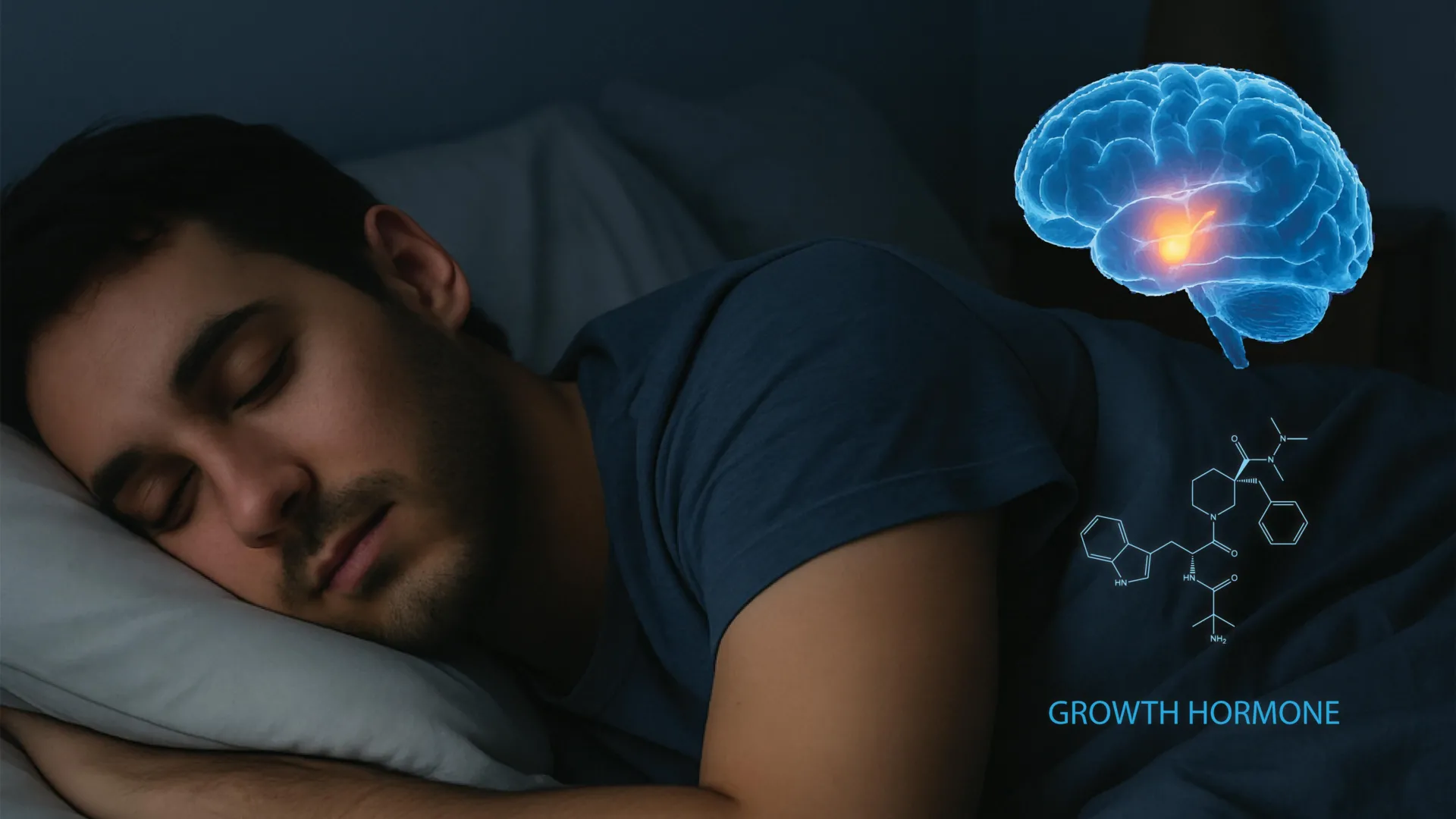
The fundamental relationship between restorative rest and physical development has long been recognized by medical science, yet the underlying biological machinery governing this connection has remained largely shrouded in mystery. While it is a well-established medical fact that a good night’s sleep triggers the release of growth hormone (GH)—a critical protein responsible for muscle synthesis, bone density, fat metabolism, and cellular repair—the precise neural pathways that command this release have only recently been mapped. Researchers at the University of California, Berkeley, have now identified the specific brain circuitry responsible for regulating growth hormone during sleep, a discovery that could redefine the clinical approach to metabolic disorders and neurodegenerative diseases.
The study, published in the prestigious journal Cell, details a sophisticated feedback system within the brain that balances hormone levels with the sleep-wake cycle. By utilizing advanced neuroimaging and optogenetic techniques, the research team has provided the first direct observation of how neural activity in the hypothalamus and the brainstem coordinates the pulsatile release of growth hormone. This breakthrough offers a blueprint for future therapeutic interventions targeting sleep-related health issues, ranging from childhood growth deficiencies to adult-onset metabolic syndromes such as Type 2 diabetes.
The Biological Significance of Growth Hormone
Growth hormone, produced by the pituitary gland, serves as a master regulator of human physiology. In children and adolescents, it is the primary driver of height and physical maturation. In adults, its role shifts toward the maintenance of lean body mass, the regulation of glucose levels, and the facilitation of lipolysis, or the breakdown of fats. Historically, athletes and fitness professionals have prioritized "deep sleep" specifically because of its association with GH release, which accelerates recovery from physical exertion and injury.
However, the consequences of GH dysregulation extend far beyond athletic performance. Chronic sleep deprivation, which affects approximately one-third of the adult population in the United States according to the Centers for Disease Control and Prevention (CDC), leads to a significant suppression of GH levels. This suppression is a contributing factor to the rising prevalence of obesity and cardiovascular disease. When the body is deprived of the GH spikes that occur during sleep, its ability to manage insulin sensitivity and fat storage is compromised, creating a physiological environment ripe for metabolic dysfunction.
Mapping the Hypothalamic Circuitry
The UC Berkeley research team, led by Yang Dan, a professor of neuroscience and molecular and cell biology, focused their investigation on the hypothalamus. This ancient region of the brain acts as a command center for the autonomic nervous system and the endocrine system. Within the hypothalamus, the researchers identified three distinct types of neurons that engage in a complex "tug-of-war" to regulate GH.
The first group consists of growth hormone-releasing hormone (GHRH) neurons, which act as the "accelerator" for hormone production. The second and third groups are variations of somatostatin neurons, which function as the "brake," suppressing the release of GH. While scientists have known about these hormones for decades, the Berkeley study is the first to show how they fluctuate in real-time according to different stages of sleep.
By recording neural activity in mice—which share similar brain structures and hormone regulation patterns with humans—the researchers observed that GHRH and somatostatin activity is not static. During non-rapid eye movement (non-REM) sleep, particularly the deep stages, somatostatin levels drop while GHRH activity increases moderately. However, the most significant activity occurs during REM sleep, where both hormone regulators increase their output, leading to a complex modulation of GH levels.
The Discovery of a Novel Feedback Loop
Perhaps the most significant finding of the study is the identification of a previously unknown feedback loop involving the locus coeruleus (LC). The locus coeruleus is a small nucleus in the brainstem responsible for synthesizing norepinephrine, the neurotransmitter that drives alertness, attention, and the "fight or flight" response.
The UC Berkeley team discovered that as growth hormone builds up in the system during sleep, it directly activates neurons in the locus coeruleus. Initially, this activation encourages the brain to transition toward wakefulness. This explains why GH levels typically peak during the first half of the night and taper off as morning approaches. However, the study also revealed a counterintuitive mechanism: if the activity in the locus coeruleus becomes excessively high, it triggers a "fail-safe" that promotes sleepiness rather than wakefulness.
"This suggests that sleep and growth hormone form a tightly balanced system," explained Daniel Silverman, a postdoctoral fellow at UC Berkeley and co-author of the study. "Too little sleep reduces growth hormone release, and too much growth hormone can in turn push the brain toward wakefulness. This balance is essential for growth, repair, and metabolic health."
Methodology and Chronology of the Research
The path to this discovery involved years of incremental progress in the laboratory of Professor Yang Dan. The research team utilized a combination of electrophysiology and optogenetics—a technique where light is used to control neurons that have been genetically sensitized to light.
- Phase One: Identifying the Players. The team first mapped the hypothalamic neurons (GHRH and somatostatin) to determine their baseline activity during wakefulness.
- Phase Two: Sleep Cycle Analysis. Because mice sleep in short bursts throughout a 24-hour period, the researchers were able to observe hundreds of transitions between wake, REM, and non-REM sleep. This provided a massive dataset that would have been impossible to gather in human subjects.
- Phase Three: Feedback Mechanism Testing. Earlier this year, Silverman published findings regarding the locus coeruleus’s role in sleepiness. The current study integrated those findings, showing that GH acts as the primary chemical signal that "dials" the LC activity up or down.
- Phase Four: Direct Recording. Unlike previous studies that relied on delayed blood tests to measure hormone levels, the Berkeley team used direct neural recording (electrodes) to see the brain’s "orders" for GH release as they happened.
Implications for Metabolic and Cardiovascular Health
The implications of this research for public health are profound. There is a well-documented correlation between shift work, sleep apnea, and chronic insomnia with the development of Type 2 diabetes. This study provides the "missing link" by showing exactly how disrupted sleep prevents the brain from executing its metabolic maintenance routine via growth hormone.
If the neural circuit for GH release can be targeted pharmacologically, it may be possible to mitigate the metabolic damage caused by sleep disorders. For instance, therapies that stabilize the activity of GHRH neurons during fragmented sleep could potentially help patients maintain healthy glucose levels and prevent the accumulation of visceral fat.
Furthermore, the connection between growth hormone and the locus coeruleus suggests that GH may play a role in daytime cognitive function. "Growth hormone not only helps you build your muscle and bones and reduce your fat tissue, but may also have cognitive benefits, promoting your overall arousal level when you wake up," noted first author Xinlu Ding. This indicates that the "brain fog" often associated with poor sleep may be a direct result of low GH levels failing to properly stimulate the locus coeruleus during the morning hours.
Neurodegeneration: Parkinson’s and Alzheimer’s Disease
The research also opens new avenues for treating neurodegenerative conditions. The locus coeruleus is one of the first brain regions to show signs of degeneration in Parkinson’s and Alzheimer’s disease. Problems in this region are linked to the sleep disturbances that often precede the motor and cognitive symptoms of these diseases by years.
By understanding how growth hormone interacts with the locus coeruleus, scientists may be able to develop neuroprotective strategies. If the GH-LC circuit can be maintained or "re-tuned" through gene therapy or specific hormonal interventions, it might be possible to slow the progression of neurodegeneration or improve the quality of life for those already diagnosed.
Silverman highlighted the potential for experimental gene therapies: "This circuit could be a novel handle to try to dial back the excitability of the locus coeruleus, which hasn’t been talked about before."
Future Directions and Collaborative Support
The study was a collaborative effort involving researchers from both UC Berkeley and Stanford University. The work was supported by the Howard Hughes Medical Institute (HHMI), where Professor Yang Dan served as an investigator, and the Pivotal Life Sciences Chancellor’s Chair fund.
The next phase of research will likely involve investigating whether these same circuits can be manipulated to treat specific growth disorders in children without the need for systemic hormone injections, which can sometimes have adverse side effects. Additionally, researchers are interested in whether the GH-LC feedback loop is affected by aging, which could explain why both sleep quality and growth hormone levels decline as people grow older.
As the scientific community continues to unravel the complexities of the human brain, the work of the Berkeley team serves as a reminder of the foundational importance of sleep. It is not merely a period of inactivity but a highly coordinated physiological event that dictates the health of nearly every system in the body. With this new map of the brain’s growth hormone circuitry, the medical field moves one step closer to personalized treatments that align our internal chemistry with the natural rhythms of rest and wakefulness.
