
 By Lina carolina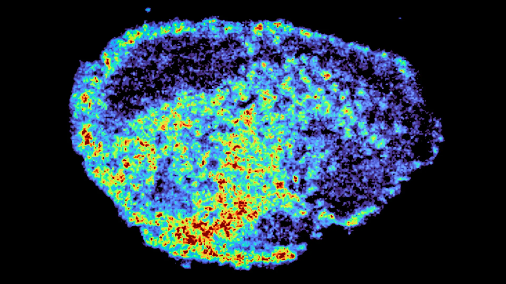
One of the most significant hurdles in modern cancer therapeutics lies in the unpredictable nature of treatment response. A groundbreaking study, published in the esteemed journal Nature Communications and spearheaded by Dr. Louise Fets at the MRC Laboratory of Medical Sciences (LMS), offers compelling new insights into why the same cancer therapy can elicit dramatically different outcomes in patients. The research zeroes in on PARP inhibitors, a vital class of targeted cancer drugs, employing sophisticated imaging techniques to meticulously track their journey within ovarian tumor samples.
The Complex Landscape of Cancer Drug Efficacy
The development of targeted cancer therapies has revolutionized patient care in recent years, offering renewed hope and improved prognoses for many. PARP inhibitors, in particular, have been transformative in the management of ovarian cancer, significantly altering treatment paradigms and enhancing survival rates for a substantial patient population. However, the persistent challenge remains: not all patients achieve the same level of benefit, and the development of treatment resistance over time is a common and concerning phenomenon. For these drugs to exert their therapeutic effect, they must attain sufficiently high concentrations within cancer cells to induce cell death. Despite the clinical success of PARP inhibitors, a fundamental gap in scientific understanding has persisted regarding the precise mechanisms that govern drug distribution within tumors and the cellular processes that influence this distribution.
This latest research illuminates a critical aspect of drug efficacy, demonstrating that successful treatment hinges not solely on whether a drug reaches a tumor, but crucially on how it disseminates both throughout the tumor microenvironment and, more granularly, within individual cancer cells. To investigate these intricate processes, the research team ingeniously utilized patient-derived ovarian tumor samples. These tumor explants, meticulously preserved to maintain their biological viability in a laboratory setting, were exposed to PARP inhibitors. This experimental design allowed scientists to directly observe and quantify drug movement within authentic human tumor tissue, providing an unparalleled level of biological relevance.
The research team employed a dual-pronged approach, leveraging cutting-edge technologies. Mass spectrometry imaging (MSI) was instrumental in generating highly detailed spatial maps, precisely delineating areas of drug accumulation within the tumor samples. Complementing this, spatial transcriptomics provided a powerful means to simultaneously examine gene expression patterns in regions exhibiting both high and low drug concentrations within the identical tissue slice. The combined power of these techniques unveiled striking variations in drug distribution. These discrepancies were not only evident when comparing different tumors from various patients but also manifested at a finer scale, within individual tumors, even when the administered drug dosage was uniform.
Dr. Zoe Hall, a senior author on the study and Associate Professor at Imperial’s Department of Metabolism, Digestion and Reproduction, emphasized the novelty of their methodology. "A novel aspect of this study was the use of mass spectrometry imaging to directly measure and visualize drug uptake in patient tumour tissue," she stated. "Through the spatial mapping of drug molecules, we could pinpoint regions of high and low drug and compare gene expression, from the same tissue slice, using spatial transcriptomics." This integrated approach allowed for an unprecedented view into the complex interplay between drug localization and cellular activity.
Lysosomes: Unveiling Hidden Drug Reservoirs
The most significant revelation from the study is the pivotal role played by lysosomes in this uneven drug distribution. Lysosomes, often referred to as the "recycling centers" of the cell, are membrane-bound organelles responsible for breaking down waste materials and cellular debris. The research found that certain PARP inhibitors are actively taken up by these lysosomes and subsequently sequestered within them, rather than dispersing uniformly throughout the cell. This intracellular sequestration creates localized "pockets" where the drug accumulates.
These lysosomes function as sophisticated, slow-release drug reservoirs. By retaining the drug and gradually releasing it, they can lead to prolonged and heightened drug exposure in specific cells while leaving other cells with significantly lower drug concentrations. Importantly, this lysosomal sequestration is not a universal phenomenon across all PARP inhibitors. The study identified that drugs such as rucaparib and niraparib are demonstrably influenced by this lysosomal mechanism, whereas others, including olaparib, appear to be less affected, suggesting distinct intracellular trafficking pathways for different drug molecules within the PARP inhibitor class.
Dr. Carmen Ramirez Moncayo, the first author of the study and a Postdoctoral Researcher at the LMS, expressed her surprise at the findings. "We were surprised to see large variability in drug accumulation at the single-cell level," she commented. "This variability was driven by the build-up of a drug in lysosomes, which are acting as reservoirs, increasing the exposure of cancer cells to drugs, by storing and releasing the drug when needed." This observation fundamentally alters our understanding of how these vital drugs achieve their therapeutic effects at the cellular level.
Implications for Precision Oncology and Future Directions
The implications of this research are profound and far-reaching, particularly for the ongoing evolution of precision oncology. PARP inhibitors are currently a cornerstone of treatment for ovarian, breast, and prostate cancers, and their therapeutic potential is actively being explored across a broad spectrum of other cancer types. A deeper comprehension of how these drugs are internalized, stored, and released within cellular compartments like lysosomes could pave the way for the development of more sophisticated and personalized treatment strategies. This, in turn, holds the promise of not only enhancing therapeutic effectiveness but also of mitigating the development of drug resistance and reducing the incidence of cancer relapse.
Dr. Louise Fets, a senior author and Head of the LMS’ Drug Transport and Tumour Metabolism Group, articulated the long-term vision. "By understanding how drugs are taken up into cells, we can understand whether this influences why cancer drugs work for some people and not for others," she explained. "Eventually, we hope to be able to study the molecular signature of a patient’s tumor to help tailor therapeutic approaches in a more personalized way." This vision aligns with the broader movement towards individualized medicine, where treatment decisions are increasingly guided by a patient’s unique biological profile.
It is crucial to acknowledge the experimental context of this study. The research was conducted using tumor tissue maintained ex vivo. In the complex in vivo environment of a living patient, drug delivery is achieved via the bloodstream. Furthermore, tumor vasculature is notoriously disorganized and heterogeneous, a factor that may further exacerbate uneven drug distribution. Future research endeavors are planned to address these complexities. These will involve the utilization of animal models, which allow for the study of drug pharmacokinetics and pharmacodynamics in a living system, and the investigation of larger patient cohorts. These studies aim to provide a more comprehensive understanding of how the interplay between drug delivery mechanisms, tumor architecture, and lysosomal sequestration influences therapeutic outcomes in clinical settings, including the challenging landscape of relapsed cancers.
Funding and Support for Cancer Research
This pivotal research was made possible through significant financial support from several key organizations dedicated to advancing cancer research. Funding was provided by the Medical Research Council (MRC) and Cancer Research UK, two leading bodies in the United Kingdom committed to combating cancer. Additional support included a PhD studentship from the Integrative Toxicology Training Partnership, administered by the MRC Toxicology Unit, fostering the next generation of scientific talent. Furthermore, a Victoria’s Secret Global Fund for Women’s Cancers Career Development Award, in partnership with Pelotonia and the American Association for Cancer Research (AACR), underscores the global commitment to addressing women’s cancers and advancing research in this critical area. The collaborative nature of this funding highlights the multifaceted effort required to unravel complex biological processes and translate discoveries into tangible patient benefits.
A Timeline of Discovery
The journey from understanding the variability in PARP inhibitor efficacy to pinpointing the role of lysosomes likely involved several stages of scientific inquiry. While the precise chronology of this specific study’s development isn’t detailed in the provided text, a typical research trajectory would involve:
- Initial Observations (Pre-2020s): Clinicians and researchers observe the variable response rates to PARP inhibitors in ovarian cancer patients, noting phenomena like primary resistance and acquired resistance. This prompts a need for deeper mechanistic understanding.
- Development of Advanced Imaging and Spatial Transcriptomics (Ongoing): The technological advancements in mass spectrometry imaging and spatial transcriptomics, which have become more accessible and sophisticated in recent years, provide the tools necessary to tackle such complex questions about drug distribution within tissue.
- Hypothesis Formulation (Early 2020s): Based on existing knowledge of cellular transport mechanisms and drug pharmacokinetics, researchers might hypothesize that intracellular sequestration, potentially within organelles like lysosomes, could explain observed variability.
- Experimental Design and Sample Acquisition (Mid-2020s): The team secures patient tumor samples and establishes the ex vivo culture system. They design experiments to deliver PARP inhibitors and employ MSI and spatial transcriptomics.
- Data Generation and Analysis (Late 2020s): Experiments are conducted, and vast amounts of data are generated. Rigorous analysis identifies the specific localization of drugs and correlates it with gene expression patterns.
- Publication and Dissemination (Early 2024 – based on Nature Communications publication date): The findings are analyzed, interpreted, and submitted for peer review, culminating in the publication of the study. This marks the official release of the new knowledge to the scientific community and the public.
- Future Research and Clinical Translation (Ongoing): The study’s findings pave the way for further investigations using in vivo models and larger patient cohorts, with the ultimate goal of translating this knowledge into improved clinical practice.
This research represents a significant step forward in demystifying the complex biological factors that influence cancer drug effectiveness, offering a glimmer of hope for more predictable and personalized cancer treatments in the future.
