
 By Nana O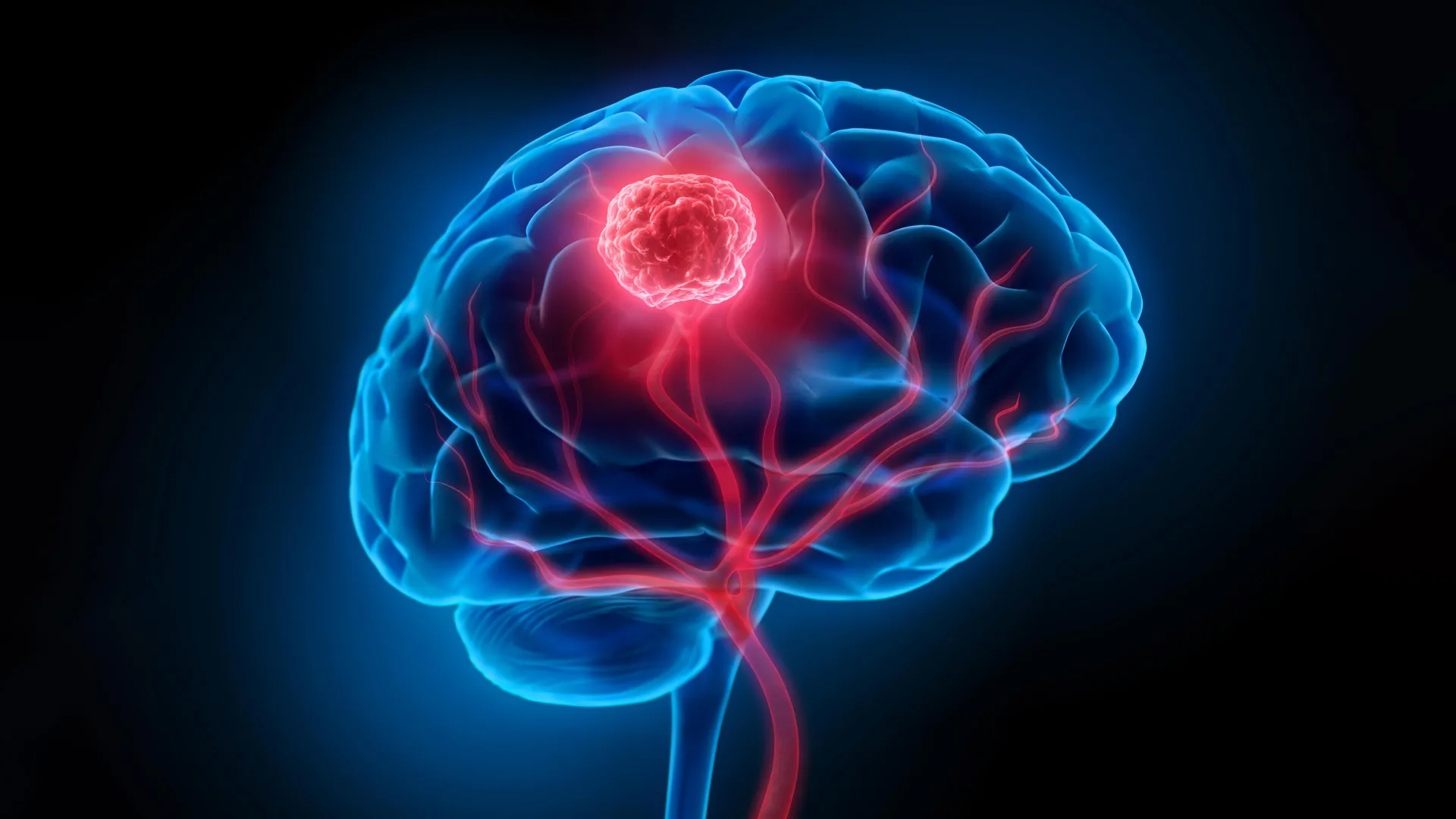
Glioblastoma, a relentless and aggressive form of brain cancer, has long represented one of medicine’s most formidable challenges. Characterized by its invasive growth into surrounding healthy brain tissue, complete surgical resection is often impossible, leaving behind microscopic cancer cells that inevitably lead to recurrence. Compounding these difficulties is the blood-brain barrier, a protective physiological shield that severely limits the efficacy of conventional drug therapies and radiotherapy. These combined obstacles contribute to a grim prognosis, with a five-year survival rate hovering at a mere 7 percent, underscoring the urgent need for innovative treatment strategies.
In a significant stride towards addressing these critical limitations, a collaborative team of researchers from the University of Technology Sydney (UTS), Harvard University, and Henan University has unveiled a novel "double-punch" nanozyme platform. This sophisticated system, detailed in the prestigious journal Science Translational Medicine, utilizes a single type of smart nanoparticle to perform two distinct yet sequential functions, aiming to revolutionize the surgical and postoperative management of glioblastoma.
The Genesis of a Dual-Action Nanotechnology
The development of this innovative platform is rooted in years of research into targeted drug delivery and advanced imaging techniques for neurological disorders. Glioblastoma’s notoriously diffuse nature has been a primary focus for oncologists and neuroscientists worldwide. Traditional surgical approaches, while vital, are often constrained by the surgeon’s ability to discern the precise boundaries of the tumor. The infiltration of cancerous cells into the delicate neural network means that aggressive removal risks irreversible neurological damage. Post-surgery, the remaining microscopic disease, invisible to the naked eye and resistant to standard treatments due to the tumor microenvironment, becomes the breeding ground for recurrence.
The research team, led by Dr. Bingyang Shi, Chair Professor of Nanomedicine at UTS, sought to create a unified solution that could overcome these sequential hurdles. "We’ve engineered a single material that does two jobs in sequence," explained Professor Shi in a recent interview. "It’s a precise guide for the surgeon during the operation, and then a targeted clean-up treatment afterward." This elegantly simple yet profound statement encapsulates the core innovation: a single, intelligent material capable of adapting its function based on external stimuli.
Engineering a Precision Tool: The Nanoparticle at its Core
At the heart of this groundbreaking system lies an exceptionally thin, two-dimensional sheet. This material, constructed atom by atom using advanced semiconductor manufacturing techniques, possesses a unique structural property that enables it to switch between two distinct roles. This meticulous atomic-level engineering is what grants the nanoparticle its remarkable versatility.
The dual functionality is activated by a specific wavelength of near-infrared light, a spectrum largely invisible to the human eye but capable of penetrating biological tissues to a useful depth. This targeted activation ensures that the nanoparticle’s functions are precisely controlled and initiated only when and where intended.
Enhancing Surgical Precision: Illuminating the Unseen
During surgical procedures, the nanoparticle platform transforms into a highly sensitive imaging agent. Professor Shi elaborated on this critical function: "During surgery, it functions as a highly sensitive imaging agent. A fluorescent dye engineered onto the sheet glows under a near-infrared wavelength invisible to the naked eye, allowing surgeons to see individual tumor cell clusters as small as 44 micrometers, a resolution beyond current clinical imaging tools."
This level of microscopic visualization is a significant advancement over existing intraoperative imaging modalities. Current techniques, while valuable, often lack the resolution to detect the smallest clusters of cancer cells that can escape macroscopic visualization. The ability to identify and target these minute infiltrations directly on the operating table could dramatically improve the completeness of tumor resection, a key determinant of patient outcomes.
Furthermore, the nanoparticles are equipped with a sophisticated targeting molecule. This molecule acts as a molecular beacon, specifically designed to bind to glioma cells. Crucially, it also facilitates the nanoparticle’s ability to overcome the formidable blood-brain barrier. This targeted accumulation ensures that the imaging agent concentrates within the tumorous tissue, minimizing off-target effects and maximizing the clarity of the imaging signal.
Postoperative Intervention: Eradicating Residual Disease
Once the visible tumor mass has been surgically removed, the same nanoparticle platform is deployed to address the insidious threat of residual microscopic cancer cells. Administered into the surgical cavity, the material is again activated by the same near-infrared light. This second activation triggers its therapeutic functions.
"After the visible tumor is removed, the same material is administered into the surgical cavity and reactivated with the same wavelength of light for postoperative phototherapy," Professor Shi explained. The mechanism of action here is multifaceted and highly effective. The platinum atoms integrated into the nanoparticle structure play a pivotal role. They catalyze a reaction with the tumor’s own hydrogen peroxide, converting it into oxygen. This process is critical because glioblastoma tumors often create a hypoxic (low-oxygen) microenvironment. This low-oxygen state is a well-known shield for cancer cells, rendering them less susceptible to conventional treatments like chemotherapy and radiotherapy. By increasing oxygen levels, the nanoparticles disrupt this protective mechanism, making any remaining cancer cells more vulnerable.
Simultaneously, the near-infrared light activates the nanoparticles to generate localized heat and reactive oxygen species. These potent agents are highly destructive to cancer cells, effectively eradicating the microscopic disease that surgery could not reach. This combined approach – altering the tumor microenvironment and directly attacking cancer cells – represents a significant departure from single-modality treatments.
Addressing the Scourge of Recurrence: Data from Preclinical Trials
The primary objective of this postoperative therapy is to combat glioblastoma’s notorious tendency for recurrence. Microscopic cancer cells that inevitably remain after surgery are the seeds of future tumor growth. The UTS, Harvard, and Henan University collaboration has demonstrated the efficacy of their nanoparticle platform in preclinical models.
In rigorous studies conducted on mouse models engineered to mimic human glioblastoma, the nanoparticle treatment significantly reduced tumor recurrence following surgery. The results were striking: every mouse treated with the nanozyme platform remained alive at the 60-day mark. In stark contrast, the control group that underwent surgery alone had an average survival of only 42 days. This substantial improvement in survival underscores the platform’s potential to fundamentally alter the trajectory of the disease.
Furthermore, comprehensive follow-up assessments in these animal models revealed no detectable neurological or motor impairments associated with the treatment. This finding is of paramount importance, as any new therapeutic strategy must not only be effective but also safe, particularly when dealing with the delicate architecture of the brain. The absence of observable side effects in the preclinical setting is a highly encouraging indicator for future human trials.
A Beacon of Hope, Yet Early Days
While the findings are undeniably promising, the research team is careful to emphasize that this technology is still in its nascent stages. The successful outcomes observed in animal models are a critical first step, but the translation to human patients requires extensive further investigation.
"The results are very encouraging, but this is still early-stage research carried out in mouse models, not in people – and that distinction is important," Professor Shi reiterated. "Its imaging and therapeutic performance will also need to be confirmed at the scale of a human brain."
The complexities of the human brain, including its size, intricate vascularization, and the variability in tumor presentation, necessitate rigorous clinical trials to validate the nanoparticle platform’s safety and efficacy in a human context. Scaling up the technology from laboratory benches to clinical application will involve overcoming significant manufacturing and regulatory hurdles.
Broader Implications and Future Directions
The potential implications of this dual-function nanoparticle platform are far-reaching. If successful in human trials, it could usher in a new era of glioblastoma management, characterized by enhanced surgical precision and a robust defense against postoperative recurrence. The ability for surgeons to "see" more of the tumor and then effectively "clean up" any residual disease offers a tangible pathway to improving patient prognoses and quality of life.
The development also signifies a broader trend in nanomedicine: the creation of multi-functional therapeutic agents. By integrating multiple capabilities into a single, intelligent system, researchers are moving away from fragmented treatment approaches towards more holistic and efficient interventions. This integrated strategy holds the promise of not only improving outcomes for glioblastoma patients but also paving the way for similar advancements in treating other complex and aggressive cancers.
The journey from laboratory discovery to clinical reality is often long and arduous, marked by rigorous testing, refinement, and regulatory approval. However, the ingenuity and promising preclinical results of this nanozyme platform provide a powerful new avenue of exploration in the relentless pursuit of a cure for glioblastoma. The scientific community will be closely watching as this innovative technology progresses, holding out hope that it may one day significantly alter the grim statistics associated with this devastating disease. The collaborative spirit demonstrated by UTS, Harvard, and Henan universities exemplifies the kind of global scientific synergy required to tackle humanity’s most pressing health challenges.
