
 By Nana O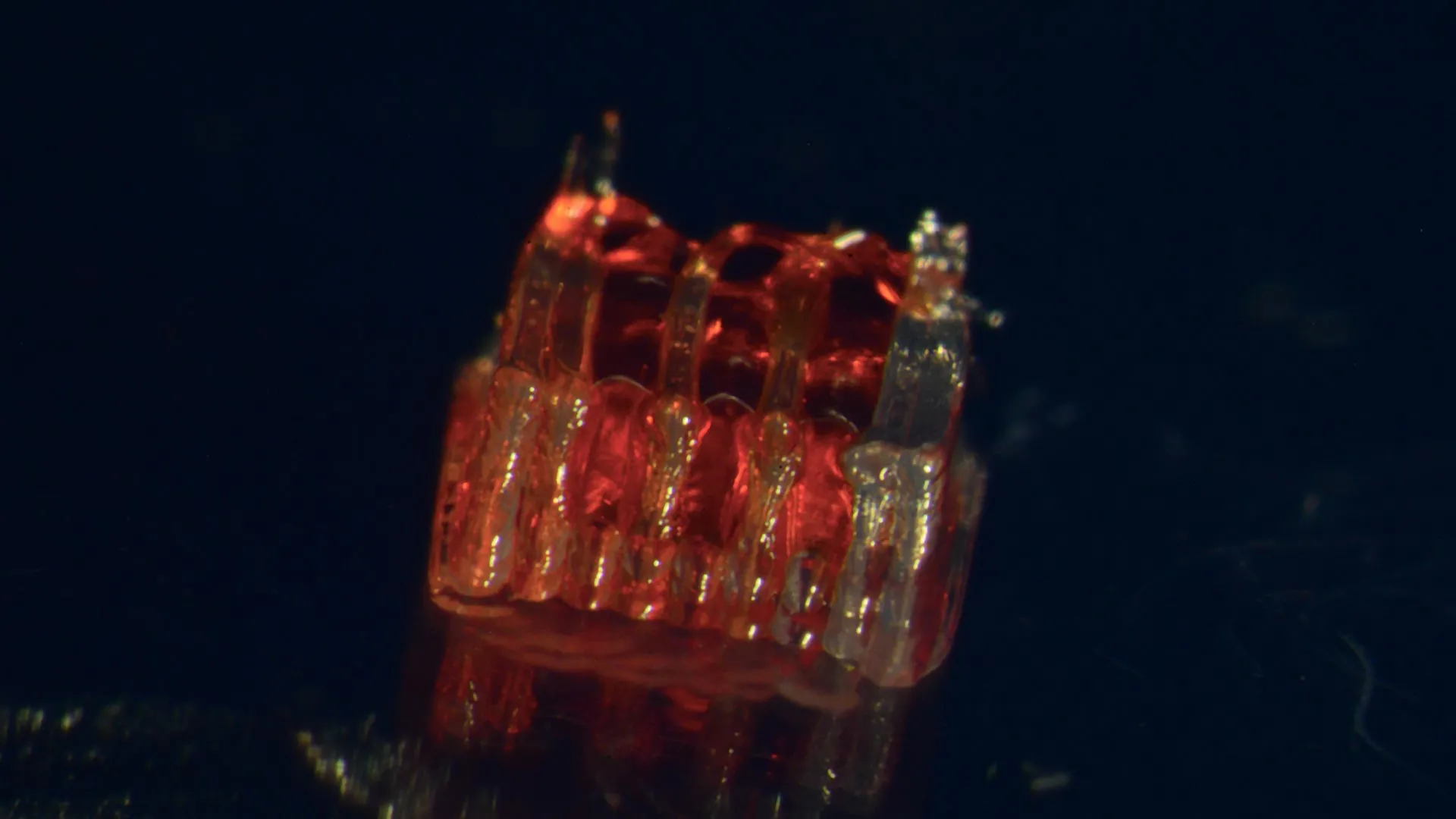
A groundbreaking advancement in regenerative medicine has emerged from the University of Minnesota Twin Cities, where a research team has successfully pioneered a novel approach to spinal cord injury recovery. For the first time, scientists have integrated 3D printing technology with stem cell biology and the creation of lab-grown tissues, offering a beacon of hope for individuals grappling with the devastating effects of spinal cord damage. This pioneering work, detailed in the prestigious peer-reviewed journal Advanced Healthcare Materials, represents a significant leap forward in the long-standing quest to repair the intricate and delicate network of the spinal cord.
The severity of spinal cord injuries cannot be overstated. According to the National Spinal Cord Injury Statistical Center, over 300,000 individuals in the United States live with the consequences of such injuries, facing challenges that often include permanent paralysis and a loss of sensation. A primary obstacle in treating these injuries lies in the inherent vulnerability of nerve cells within the spinal cord. Once damaged or destroyed, these neurons possess a limited capacity for natural regeneration, and the severed nerve fibers struggle to bridge the gap created by the injury. This fundamental biological limitation has rendered most spinal cord injuries irreversible, leaving patients with few options for functional restoration. The University of Minnesota team’s research directly confronts this critical challenge by devising a strategy to guide and facilitate nerve regrowth across the injured site.
Crafting a Neural Relay System: The 3D-Printed Scaffold
At the core of this innovative solution is a meticulously designed 3D-printed framework, referred to as an organoid scaffold. This scaffold is not merely a structural component; it is engineered with microscopic channels, a feature crucial for its biological function. These channels serve as precise conduits, guiding the growth and organization of specialized cells. The researchers then populate these channels with regionally specific spinal neural progenitor cells (sNPCs). These sNPCs are derived from human adult stem cells, granting them the remarkable plasticity to differentiate into various specialized cell types, including the neurons essential for spinal cord function.
Dr. Guebum Han, a former postdoctoral researcher in mechanical engineering at the University of Minnesota and the study’s lead author, elaborated on the ingenious design. "We use the 3D printed channels of the scaffold to direct the growth of the stem cells, which ensures the new nerve fibers grow in the desired way," Dr. Han explained. "This method creates a relay system that when placed in the spinal cord bypasses the damaged area." This "relay system" is a critical conceptual breakthrough, envisioning the scaffold as a bridge that allows neural signals to circumvent the site of injury, thereby restoring communication pathways. The precise control offered by the 3D printing technology allows for an unprecedented level of organization, mimicking the natural architecture of the spinal cord to an extent previously unattainable.
A Glimpse into the Future: Pre-Clinical Success in Animal Models
The efficacy of this novel approach was rigorously tested through pre-clinical trials involving rats with completely severed spinal cords. In a pivotal experiment, these meticulously engineered scaffolds, populated with sNPCs, were transplanted into the injured spinal cords of the animals. The results were profoundly encouraging. The transplanted cells not only survived but also successfully differentiated into mature neurons. Crucially, these newly formed nerve fibers exhibited robust growth, extending in both directions – rostral (towards the head) and caudal (towards the tail) – to establish new synaptic connections with the host’s existing neural circuitry.
The seamless integration of these new nerve cells into the host spinal cord tissue was a key observation. Over time, the researchers witnessed a remarkable phenomenon: the new neural network began to function, facilitating the transmission of signals across the previously irreparable gap. This integration translated into significant functional recovery in the rats. While the specifics of the functional recovery were not detailed in the initial report, the implication of restored motor or sensory function in an animal model of complete spinal cord transection is a powerful testament to the potential of this technology. This success in a severe injury model offers a tangible demonstration of the scaffold’s ability to promote meaningful neural regeneration and functional restoration.
The Dawn of a New Era in Spinal Cord Injury Research
The implications of this research extend far beyond the laboratory. Ann Parr, a professor of neurosurgery at the University of Minnesota and a senior figure in the study, expressed palpable enthusiasm for the findings. "Regenerative medicine has brought about a new era in spinal cord injury research," Professor Parr stated. "Our laboratory is excited to explore the future potential of our ‘mini spinal cords’ for clinical translation." The term "mini spinal cords" aptly captures the essence of the engineered tissue – a compact, functional unit designed to mimic and restore the capabilities of the native spinal cord. This sentiment underscores the team’s commitment to translating their laboratory breakthroughs into tangible treatments for human patients.
While acknowledging that the research is still in its nascent stages, the development represents a significant paradigm shift in how spinal cord injuries are approached. The combination of advanced manufacturing (3D printing), sophisticated cell biology (stem cell differentiation), and tissue engineering offers a multi-faceted strategy that addresses the complex challenges of neural repair. The team’s immediate goal is to scale up the production of these organoid scaffolds and to refine the techniques involved, paving the way for future clinical trials in humans. This iterative process of research, development, and testing is essential for ensuring the safety and efficacy of any new medical intervention.
Building on a Foundation of Collaboration and Funding
The success of this ambitious project is a testament to the collaborative spirit of the scientific community and the critical role of dedicated funding. The research team comprised a diverse group of experts from various departments at the University of Minnesota, including Mechanical Engineering, Neurosurgery, and Neuroscience. Key contributors included Hyunjun Kim and Michael McAlpine from Mechanical Engineering; Nicolas S. Lavoie, Nandadevi Patil, and Olivia G. Korenfeld from Neurosurgery; and Manuel Esguerra from Neuroscience. The project also benefited from the expertise of Daeha Joung from the Department of Physics at Virginia Commonwealth University, highlighting the interdisciplinary nature of modern scientific inquiry.
This groundbreaking work was made possible through the generous support of several key funding bodies. The National Institutes of Health, a cornerstone of biomedical research funding in the United States, provided crucial financial backing. Additionally, the State of Minnesota’s Spinal Cord Injury and Traumatic Brain Injury Research Grant Program and the Spinal Cord Society offered vital support, underscoring a shared commitment to alleviating the burden of spinal cord injuries. The comprehensive financial and intellectual investment in this research project signals a strong belief in its transformative potential.
The Road Ahead: Challenges and Opportunities
The path from laboratory discovery to clinical application is often long and fraught with challenges. However, the initial success of this 3D-printed organoid scaffold offers a compelling new avenue of hope for the millions affected by spinal cord injuries. The ability to precisely control cellular organization and guide nerve regeneration represents a significant departure from previous treatment strategies, which have largely focused on mitigating secondary damage rather than actively promoting repair.
Future research will undoubtedly focus on several critical areas. Further pre-clinical studies will be essential to thoroughly assess the long-term safety and efficacy of the transplanted scaffolds, including potential immune responses and the stability of the regenerated neural connections. Optimizing the differentiation and integration of sNPCs within the scaffold will also be a key area of investigation, aiming to maximize the functional output of the engineered tissue. Furthermore, the development of minimally invasive surgical techniques for implantation will be crucial for facilitating widespread clinical adoption.
The broader implications of this research are profound. Beyond spinal cord injuries, the principles of 3D-printed organoid scaffolds could potentially be applied to other neurological conditions characterized by nerve damage, such as stroke, traumatic brain injury, and neurodegenerative diseases. The ability to create complex, functional biological structures on demand opens up a vast landscape of possibilities for regenerative medicine and personalized therapies. As this research continues to evolve, it holds the promise of transforming the lives of countless individuals and redefining the boundaries of what is medically possible. The full details of this pioneering study can be found in the paper entitled, "3D-Printed Scaffolds Promote Enhanced Spinal Organoid Formation for Use in Spinal Cord Injury," published on the Advanced Healthcare Materials website.
