
 By George Ongkojoyo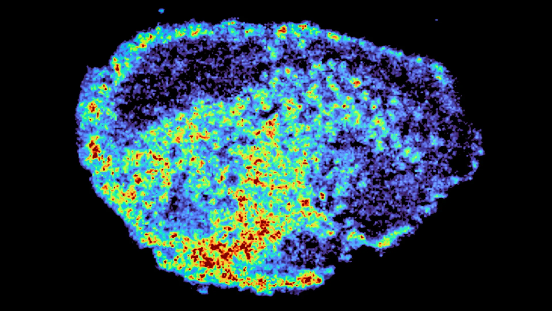
The landscape of modern oncology is defined by a persistent and frustrating paradox: therapies that prove life-saving for one patient often fail entirely for another, even when the clinical presentation of the disease appears identical. This variability in drug response remains one of the most significant hurdles in achieving consistent outcomes in cancer care. A groundbreaking study published in the journal Nature Communications has shed new light on this phenomenon, revealing that the internal architecture of cancer cells—specifically small organelles known as lysosomes—plays a decisive role in how drugs are distributed and retained within a tumor. Led by Dr. Louise Fets at the MRC Laboratory of Medical Sciences (LMS), the research focused on a vital class of targeted therapies known as PARP inhibitors, uncovering how these drugs can become trapped within cellular "recycling centers," acting as hidden reservoirs that influence treatment efficacy.
The Evolution and Impact of PARP Inhibitors
To understand the significance of this discovery, one must first look at the role of Poly (ADP-ribose) polymerase (PARP) inhibitors in contemporary cancer treatment. Since their introduction, PARP inhibitors have revolutionized the management of several malignancies, most notably ovarian, breast, prostate, and pancreatic cancers. These drugs operate on the principle of "synthetic lethality." In patients with mutations in the BRCA1 or BRCA2 genes, the cells already have a compromised ability to repair double-strand DNA breaks. By inhibiting the PARP enzyme, which is responsible for repairing single-strand breaks, the drugs force the cancer cells into a state where they can no longer repair their DNA at all, leading to programmed cell death.
For ovarian cancer patients, PARP inhibitors like olaparib, rucaparib, and niraparib have extended progression-free survival significantly. However, the clinical reality is far from perfect. While some patients experience dramatic remissions, others exhibit primary resistance, where the drug never works, or acquired resistance, where the tumor eventually learns to bypass the drug’s effects. Until now, much of the research into resistance has focused on genetic mutations that alter DNA repair pathways. The study by Dr. Fets and her colleagues shifts the focus to the physical distribution of the drug itself, suggesting that if the medicine cannot reach its intended target within the cell nucleus in sufficient concentrations, its genetic impact is moot.
Methodology: Mapping the Intracellular Journey
The research team sought to bridge the gap between systemic drug delivery and cellular-level uptake. In a typical clinical setting, a drug is administered orally or intravenously, travels through the bloodstream, and eventually infiltrates the tumor mass. However, once inside the tumor, the drug’s journey is far from over. It must navigate a complex microenvironment and penetrate individual cell membranes to reach the molecular machinery it is designed to inhibit.
To observe this process in high resolution, the researchers utilized "explants"—thin, viable slices of ovarian tumors harvested from patients. By maintaining these tissues in a laboratory environment that mimics the human body, the team could treat the samples with PARP inhibitors and track their movement through real human tissue. This approach is significantly more sophisticated than using traditional cell cultures, as it preserves the structural complexity and cellular diversity of a real tumor.
The study employed a dual-track imaging strategy to visualize drug distribution. First, the team used mass spectrometry imaging (MSI), a powerful tool that allows scientists to create heat maps showing the exact concentration of drug molecules across a tissue sample. This was paired with spatial transcriptomics, an emerging technology that measures gene activity in specific locations. by overlaying these two maps, the researchers could compare the gene expression profiles of areas with high drug accumulation against areas where the drug was largely absent.
The Lysosomal Reservoir Discovery
The most striking finding of the study was the uneven distribution of drugs within the tumor tissue. Even when exposed to identical doses, different regions of the same tumor showed vastly different levels of drug uptake. The primary driver of this disparity was identified as the lysosome.
Lysosomes are often described as the "stomachs" or "recycling centers" of the cell. They are acidic organelles filled with enzymes that break down waste products and cellular debris. The researchers discovered that certain PARP inhibitors—specifically rucaparib and niraparib—are "lysosomotropic." Because of their chemical properties as weak bases, they are naturally drawn into the acidic environment of the lysosome. Once inside, they become protonated (gain a hydrogen ion), which traps them within the organelle membrane.
This sequestration creates internal "pockets" where the drug accumulates at much higher levels than in the rest of the cell. Dr. Carmen Ramirez Moncayo, the study’s first author and a Postdoctoral Researcher at the LMS, noted the team’s surprise at the degree of single-cell variability. The lysosomes were not merely destroying the drug; they were acting as slow-release reservoirs. These organelles store the drug and release it gradually over time, which can actually increase the duration of exposure for certain cancer cells. However, this also means that the drug is not immediately available to the nucleus, where the PARP enzymes reside, potentially delaying or altering the therapeutic effect.
Comparative Analysis of PARP Inhibitor Behavior
One of the study’s critical contributions is the distinction it draws between different drugs within the same class. While rucaparib and niraparib showed significant lysosomal accumulation, olaparib did not. This chemical nuance explains why patients might respond differently to one PARP inhibitor versus another, despite their similar primary mechanisms of action.
This finding aligns with known pharmacokinetic data but provides a much-needed visual and biological explanation. The ability of a drug to be stored and slowly released by lysosomes could be a double-edged sword. On one hand, it might provide a "buffer" that keeps drug levels stable between doses. On the other hand, if too much of the drug is sequestered away from the DNA repair machinery, it could allow the cancer cell to survive despite treatment. This variability was observed not only between different drugs but also between different patients, suggesting that the "lysosomal load" or the acidity of a patient’s tumor cells could be a predictor of how well they will respond to specific therapies.
Professional Perspectives and Industry Reaction
The implications of this research have resonated throughout the oncological community. Dr. Zoe Hall, senior author and Associate Professor at Imperial College London’s Department of Metabolism, Digestion and Reproduction, emphasized the novelty of the spatial mapping approach. "Through the spatial mapping of drug molecules, we could pinpoint regions of high and low drug and compare gene expression from the same tissue slice," Dr. Hall stated. This ability to link drug concentration directly to biological response at a microscopic level provides a blueprint for future drug development.
Dr. Louise Fets, Head of the LMS’ Drug Transport and Tumor Metabolism Group, highlighted the potential for personalized medicine. "By understanding how drugs are taken up into cells, we can understand whether this influences why cancer drugs work for some people and not for others," she explained. The ultimate goal, according to Dr. Fets, is to develop a "molecular signature" for each patient’s tumor. If a clinician knows a tumor has high lysosomal activity, they might choose a non-lysosomotropic drug like olaparib over niraparib to ensure the medication reaches the cell nucleus effectively.
Broader Implications and Future Research Directions
The study opens several new avenues for clinical investigation. While the current research was conducted on tissue explants, the next phase involves animal models to see how systemic factors—such as blood flow and disorganized tumor vasculature—interact with lysosomal storage. In a living patient, the delivery of the drug is often hampered by the chaotic growth of tumor blood vessels, which creates "dead zones" where medicine cannot reach. Combining the challenges of poor delivery with the challenges of lysosomal sequestration could explain why some aggressive cancers are so difficult to treat.
Furthermore, there is the question of drug resistance in relapsed cancers. If lysosomes are acting as reservoirs, they might contribute to the survival of "persister cells"—small populations of cancer cells that survive initial treatment and eventually lead to a recurrence. Future studies will likely investigate whether drugs that alter lysosomal pH could be used in combination with PARP inhibitors to "unlock" the trapped medicine and enhance its lethality.
The research also has implications beyond PARP inhibitors. Many other cancer drugs, including certain types of chemotherapy and targeted kinase inhibitors, have chemical structures that make them susceptible to lysosomal trapping. The methodology developed by the LMS and Imperial College team could be applied to a wide range of therapies to map their distribution and optimize their design.
Conclusion and Funding Acknowledgments
This research represents a significant step forward in our understanding of the "pharmacological microenvironment." It moves the conversation beyond just "is the drug in the tumor?" to "where is the drug inside the cell?" As the medical community moves toward a more personalized approach to oncology, factors like organelle-level drug distribution will become increasingly vital in choosing the right treatment for the right patient.
The study was a collaborative effort supported by a diverse range of prestigious organizations. Funding was provided by the Medical Research Council (MRC) and Cancer Research UK. Additional support came from a PhD studentship via the Integrative Toxicology Training Partnership (administered by the MRC Toxicology Unit) and a Victoria’s Secret Global Fund for Women’s Cancers Career Development Award, in partnership with Pelotonia and the American Association for Cancer Research (AACR).
As researchers continue to decode the complex relationship between cellular anatomy and drug efficacy, the hope is that the high variability in cancer treatment outcomes will finally begin to narrow, leading to more predictable and successful interventions for patients worldwide.
