
 By Nana O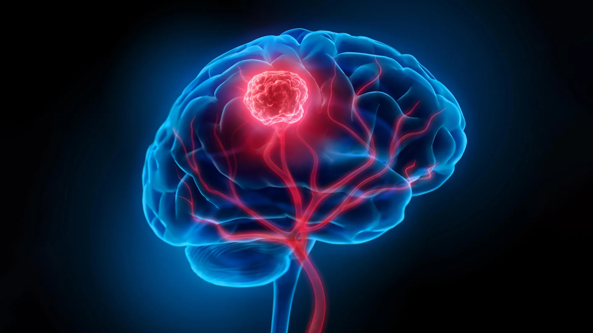
Glioblastoma, a formidable and relentlessly aggressive form of brain cancer, has long presented an insurmountable challenge to medical science. Characterized by its rapid growth and invasive nature, glioblastoma infiltrates surrounding healthy brain tissue, making complete surgical eradication a perilous undertaking. Surgeons are compelled to navigate a delicate balance, aiming to excise the tumor while meticulously preserving vital neurological functions. This inherent difficulty is compounded by the formidable blood-brain barrier, a biological shield that significantly impedes the efficacy of conventional treatments like chemotherapy and radiotherapy, limiting their ability to penetrate the tumor site. Consequently, the prognosis for patients diagnosed with glioblastoma remains grim, with a stark five-year survival rate hovering around a mere 7 percent, underscoring the urgent need for innovative therapeutic strategies.
However, a groundbreaking development from a collaborative effort between researchers at the University of Technology Sydney (UTS), Harvard University, and Henan University may herald a new era in glioblastoma treatment. Their pioneering work, published in the prestigious journal Science Translational Medicine, introduces a novel ‘double-punch’ nanozyme platform, a sophisticated system of intelligent nanoparticles engineered to address both the surgical and therapeutic challenges posed by this devastating disease.
A Dual-Action Nanoparticle System: Precision Imaging and Targeted Therapy
At the heart of this revolutionary approach lies an exceptionally thin, two-dimensional sheet composed of individual atoms meticulously arranged using advanced semiconductor manufacturing techniques. This unique atomic-level architecture grants the material an extraordinary ability to seamlessly transition between two distinct, yet complementary, functions: precise intraoperative imaging and targeted postoperative phototherapy. Crucially, both of these functions are activated by the same wavelength of near-infrared light, a non-invasive and readily administrable energy source.
Dr. Bingyang Shi, Chair Professor of Nanomedicine at UTS’s School of Electrical, Mechanical and Biomedical Engineering, explained the ingenious design. "We’ve engineered a single material that does two jobs in sequence," he stated. "It’s a precise guide for the surgeon during the operation, and then a targeted clean-up treatment afterward." This sequential functionality represents a significant leap forward, offering a comprehensive solution within a single, integrated system.
Enhancing Surgical Precision: Visualizing Microscopic Tumors
The initial application of this nanozyme platform during surgery is as a highly sensitive imaging agent. A specially engineered fluorescent dye, embedded within the nanoparticle sheet, emits a glow when exposed to near-infrared light, a wavelength invisible to the human eye. This fluorescence allows surgeons to visualize even the smallest clusters of glioblastoma cells, as diminutive as 44 micrometers. This level of resolution far surpasses the capabilities of current clinical imaging technologies, enabling surgeons to identify and remove tumor margins with unprecedented accuracy.
Furthermore, the nanoparticles are equipped with a targeting molecule that facilitates their passage across the blood-brain barrier. This crucial feature ensures that the nanoparticles selectively accumulate within glioma cells, minimizing off-target effects and maximizing their presence at the tumor site. This targeted accumulation is vital for both accurate imaging and subsequent therapeutic intervention. By providing a clear visual map of the cancerous infiltration, the nanozyme platform empowers surgeons to resect as much of the tumor as safely possible, significantly reducing the likelihood of leaving residual disease behind.
Post-Operative Clean-Up: Eliminating Residual Cancer Cells
Following the visible tumor’s removal, the nanozyme platform is reintroduced into the surgical cavity. Upon reactivation with the same near-infrared light, the nanoparticles initiate their therapeutic phase. The platinum atoms within the nanostructure play a critical role by converting the tumor’s endogenous hydrogen peroxide into oxygen. This process is pivotal in counteracting the hypoxic (low-oxygen) microenvironment that typically shields glioblastoma cells from treatment, rendering them more resistant to conventional therapies.
Simultaneously, the applied near-infrared light generates heat and reactive oxygen species (ROS). These localized thermal effects and chemically reactive molecules work in concert to destroy any microscopic cancer cells that may have evaded surgical removal. This ‘clean-up’ phase is arguably the most critical aspect of the technology, directly addressing the primary driver of glioblastoma recurrence: the persistent presence of microscopic, undetectable cancer cells.
Addressing the Specter of Recurrence: A Critical Challenge in Glioblastoma Management
The tenacious ability of glioblastoma to recur after initial treatment is a grim reality for patients and a significant hurdle for oncologists. Even with meticulous surgical resection, microscopic cancer cells can persist within the brain’s complex architecture, lying dormant until they proliferate and initiate a new tumor. This insidious recurrence is often responsible for the poor long-term survival rates associated with the disease.
The dual-function nanoparticle platform is specifically designed to confront this challenge head-on. By enhancing surgical visualization and then providing a targeted therapeutic intervention to eliminate residual cells, the technology offers a comprehensive strategy to disrupt the cycle of recurrence.
Promising Preclinical Results: A Glimmer of Hope in Animal Models
The efficacy of this innovative approach has been rigorously tested in preclinical studies utilizing mouse models of glioblastoma. The results have been remarkably encouraging. In mice treated with the nanoparticle platform following surgery, tumor recurrence was significantly reduced. Notably, all treated mice remained alive at the 60-day follow-up mark, a stark contrast to the control group that received surgery alone, where the average survival was only 42 days.
Crucially, extensive follow-up testing in these animal models revealed no detectable neurological or motor impairments associated with the nanoparticle treatment. This finding is of paramount importance, as any new therapeutic intervention must demonstrate a favorable safety profile, especially when targeting the delicate environment of the brain. The absence of adverse neurological effects suggests that the nanozyme platform can achieve its therapeutic goals without compromising cognitive or motor functions, a critical consideration for improving the quality of life for glioblastoma patients.
The Road Ahead: From Bench to Bedside
Despite the overwhelmingly positive preclinical outcomes, the researchers involved are keen to temper expectations and emphasize that this technology is still in its nascent stages of development. "The results are very encouraging, but this is still early-stage research carried out in mouse models, not in people – and that distinction is important," Professor Shi cautioned. The transition from animal models to human clinical trials is a complex and lengthy process, requiring extensive validation and rigorous safety testing.
The next critical phase will involve scaling up the technology to match the anatomical complexities of the human brain. The imaging and therapeutic performance of the nanoparticles will need to be re-evaluated and confirmed in larger biological systems. This includes assessing how the nanoparticles distribute, function, and are cleared from the human body, as well as further refining the delivery mechanisms and light activation protocols for optimal patient outcomes.
Broader Implications and Future Directions
If this technology successfully navigates the path through clinical trials and gains regulatory approval, it could fundamentally alter the landscape of glioblastoma treatment. The ability for surgeons to "see more of the tumor during an operation and treat more of what’s left behind afterward" represents a profound advancement. This dual-action approach has the potential to significantly improve patient survival rates and, perhaps more importantly, enhance their quality of life by minimizing the debilitating effects of tumor recurrence.
The implications extend beyond glioblastoma. The underlying principles of this dual-function nanoparticle platform could potentially be adapted to target other aggressive and invasive cancers, particularly those that are difficult to resect surgically or are resistant to conventional therapies. The ability to combine precise imaging with targeted therapeutic delivery, all activated by a single, non-invasive stimulus, opens up exciting avenues for future research and development in nanomedicine.
The collaborative spirit that has driven this innovation, bringing together expertise from diverse institutions across continents, highlights the power of interdisciplinary research in tackling some of the most pressing challenges in healthcare. As this promising technology continues its journey from the laboratory to potential clinical application, it offers a tangible beacon of hope for patients and families grappling with the devastating reality of glioblastoma. The scientific community will be closely watching as this remarkable nanozyme platform unfolds, potentially rewriting the prognosis for one of the most aggressive forms of cancer known to humankind.
