
 By Billy Candra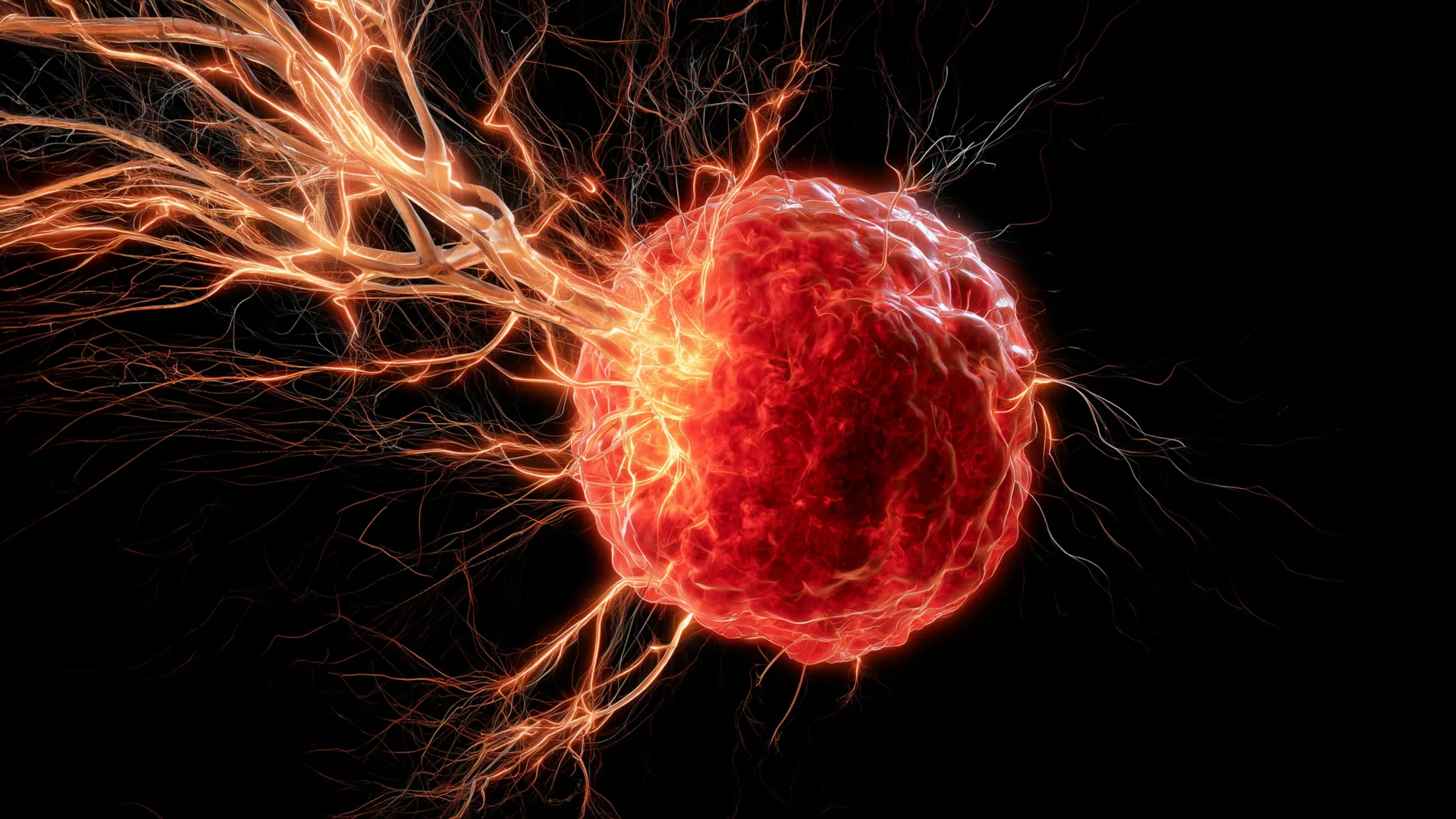
New research from the University of Oklahoma has revealed a critical and previously underappreciated pathway by which an aggressive form of breast cancer, triple-negative breast cancer (TNBC), manipulates the body’s own immune system to facilitate its growth and potential resistance to therapy. The study, published in the esteemed journal Cell Death & Differentiation, details how specific immune cells, known as macrophages, are recruited by tumors and subsequently release a protein that actively draws nerves into the cancerous mass, creating a unique microenvironment that may contribute to disease progression. This discovery sheds new light on the intricate interplay between cancer cells, immune components, and the nervous system, opening promising avenues for novel therapeutic strategies.
For decades, scientists have observed the presence of extensive nerve networks within many solid tumors, a phenomenon known as tumor innervation or neurogenesis. However, the precise mechanisms by which these nerves are recruited into the tumor microenvironment – the complex ecosystem surrounding and within a tumor – have remained largely enigmatic. This new study provides a compelling explanation, specifically for triple-negative breast cancer, a particularly challenging subtype due to its aggressive nature and limited targeted treatment options.
The Enigma of Triple-Negative Breast Cancer
Triple-negative breast cancer (TNBC) represents a significant clinical challenge in oncology. Accounting for approximately 10-15% of all breast cancers, it is characterized by the absence of estrogen receptors (ER), progesterone receptors (PR), and human epidermal growth factor receptor 2 (HER2) overexpression. This "triple-negative" status means that TNBC does not respond to hormone therapies (which target ER/PR) or HER2-targeted therapies like trastuzumab. Consequently, standard treatment for TNBC relies primarily on chemotherapy, often combined with surgery and radiation. Despite advancements, TNBC is associated with a higher risk of recurrence and metastasis, and a poorer prognosis compared to other breast cancer subtypes, particularly in the early stages and for younger women. The aggressive biological behavior of TNBC underscores an urgent need for novel therapeutic approaches that can effectively target its unique characteristics.
The tumor microenvironment (TME) is now recognized as a critical determinant of cancer progression, metastasis, and therapeutic response. Far from being a mere collection of malignant cells, a tumor exists within a dynamic ecosystem comprising various cell types, including immune cells, fibroblasts, endothelial cells (forming blood vessels), and an extracellular matrix. This intricate network actively supports tumor growth, survival, and evasion of immune surveillance. Among the diverse cellular inhabitants of the TME, macrophages, a type of immune cell, play a particularly paradoxical role.
Macrophages: From Defenders to Accomplices
Macrophages are large phagocytic immune cells that originate from monocytes and are found in virtually all tissues. Their primary physiological roles include clearing cellular debris, orchestrating tissue repair, and presenting antigens to initiate adaptive immune responses against pathogens. In the context of cancer, however, macrophages often undergo a phenotypic shift, becoming "tumor-associated macrophages" (TAMs) that promote tumor growth, angiogenesis (formation of new blood vessels), immune suppression, and metastasis. This duality makes them a complex target for therapeutic intervention.
The University of Oklahoma research team, led by Dr. Maureen Cox, an assistant professor in the Department of Microbiology and Immunology at the OU College of Medicine and a research member of OU Health Stephenson Cancer Center, made a pivotal discovery regarding these macrophages in TNBC. They found that TNBC tumors actively recruit macrophages into their core. Once within the tumor microenvironment, these macrophages do not perform their typical anti-cancer functions; instead, they become key orchestrators in drawing nerves into the tumor.
The Role of Brain-Derived Neurotrophic Factor (BDNF)
The mechanism hinges on a specific protein released by these tumor-associated macrophages: brain-derived neurotrophic factor (BDNF). BDNF is a member of the neurotrophin family, widely recognized for its crucial role in the central and peripheral nervous systems. In healthy neurological contexts, BDNF supports the survival, growth, and differentiation of neurons, playing a vital part in learning, memory, and overall brain plasticity. However, in the context of TNBC, the researchers discovered that tumors exploit this powerful biological signal for their own benefit.
"Macrophages are the critical source for drawing nerves into the tumor," stated Dr. Cox. "Although macrophages typically play a positive role in the body, they are facilitating a negative function in this scenario of breast cancer." The BDNF released by these recruited macrophages acts as a potent chemoattractant, signaling nearby nerves to sprout and grow towards, and ultimately infiltrate, the cancerous mass. This process of tumor neurogenesis, or the growth of new nerves within a tumor, is increasingly recognized as a significant factor in cancer progression, influencing everything from tumor initiation to metastasis and pain perception.
Chronology of Discovery and Experimental Validation
The journey to this discovery likely followed a meticulous scientific trajectory:
- Initial Observations: Long-standing recognition of nerve presence within various solid tumors, including breast cancer.
- Hypothesis Formulation: Postulating that tumors actively recruit nerves rather than merely being passively infiltrated, and that immune cells might play a role in this recruitment.
- Experimental Design (Pre-clinical Models): The team initiated studies using preclinical mouse models of triple-negative breast cancer. These models are crucial for understanding disease mechanisms and testing potential therapies in a controlled environment.
- Identification of Key Players: Through rigorous cellular and molecular analyses, the researchers identified macrophages as central to the nerve recruitment process within the tumor microenvironment.
- Discovery of BDNF Link: Subsequent investigations pinpointed BDNF as the specific molecular signal released by macrophages that drives nerve growth towards the tumor.
- Therapeutic Intervention Testing: With the mechanism identified, the team moved to test interventions. They utilized a pharmacological agent specifically designed to block BDNF signaling. This drug, already available on the market, presented an immediate translational advantage.
- Observation of Efficacy: In the mouse models, blocking BDNF signaling effectively prevented nerves from growing into the tumors. Critically, this intervention also resulted in a significant reduction in tumor growth, underscoring the functional importance of tumor innervation in disease progression.
- Human Data Validation: To ensure the relevance of their findings to human patients, the researchers analyzed clinical data from individuals diagnosed with triple-negative breast cancer. They found a compelling correlation: tumors with higher levels of macrophages and BDNF were statistically linked with poorer patient survival outcomes. This crucial step provided strong evidence that the biological mechanism observed in mice is highly pertinent to human disease.
This systematic approach, moving from fundamental biological observation to mechanistic elucidation, preclinical validation, and finally, correlation with human patient data, exemplifies robust translational research.
Blocking the Signal: A Promising Therapeutic Avenue
The discovery that inhibiting BDNF signaling can slow tumor growth in mice offers a compelling new therapeutic strategy. Instead of solely focusing on cytotoxic approaches to destroy cancer cells, future therapies might target the intricate signaling pathways within the tumor microenvironment that support cancer’s aggressive behavior. This represents a paradigm shift in oncology, moving towards therapies that disarm the tumor’s support system rather than directly attacking the cancer cells alone.
"It looks really promising that we can use this drug, which is already on the market, to target BDNF," Dr. Cox enthusiastically noted. The potential to repurpose an existing drug to address a novel cancer mechanism is particularly exciting, as it could significantly accelerate the timeline for clinical trials and patient access, bypassing many of the lengthy and costly development stages associated with entirely new compounds.
Beyond directly hindering tumor growth, the researchers hypothesize that blocking nerve infiltration could have an additional, profound benefit: boosting the anti-tumor immune response. "We believe that the nerves are immunosuppressive, so if we can stop the nerves from growing in the first place, maybe we can boost the immune response to help fight the cancer," Dr. Cox explained. Nerves within the tumor microenvironment are increasingly implicated in creating an immunosuppressive milieu, making it harder for the body’s own immune cells to recognize and eliminate cancer cells. By disrupting this nerve-mediated immunosuppression, BDNF blockade could potentially render TNBC tumors more susceptible to immunotherapies, which have shown limited efficacy in many TNBC cases.
Broader Implications and Future Directions
The implications of this research extend beyond triple-negative breast cancer. The mechanism of immune cell-driven nerve recruitment could be a generalizable feature of other aggressive solid tumors known for extensive innervation and poor prognoses. The research team is already exploring this possibility.
"Ultimately, we want to turn the anti-tumor immunity back on in cancer patients so their own immune systems can reject the tumors," Dr. Cox emphasized. To achieve this overarching goal, the team has outlined several critical next steps:
- Deciphering Nerve Contribution: A deeper understanding of exactly how nerves contribute to tumor growth is crucial. Some evidence suggests that nerves might stimulate the formation of new blood vessels (angiogenesis), providing tumors with essential oxygen and nutrients. Other research indicates that cancer cells might utilize nerves as migratory highways to leave the primary tumor site and metastasize to distant organs, a process known as perineural invasion. Elucidating these precise roles will further refine therapeutic strategies.
- Testing in Other Cancers: The researchers plan to test the same BDNF-blocking intervention in high-grade ovarian cancer, another aggressive malignancy that presents significant treatment challenges and is often characterized by extensive nerve infiltration. Success in ovarian cancer would underscore the broad applicability of this therapeutic approach.
- Combination Therapies: Exploring the potential for combining BDNF blockade with existing chemotherapies or immunotherapies is a logical next step. Synergistic effects could lead to more potent and durable responses in patients.
- Biomarker Identification: Identifying reliable biomarkers that predict which patients are most likely to benefit from BDNF-targeted therapy would be essential for personalized medicine approaches.
This research aligns with a growing body of evidence highlighting the nervous system’s complex and often detrimental role in cancer progression. For instance, studies have shown that denervation (surgical removal of nerves) can suppress tumor growth in various models, including prostate and pancreatic cancer. The University of Oklahoma study provides a novel molecular pathway for this neuro-oncological interaction, specifically implicating the immune system as an intermediary.
The potential to repurpose an existing drug to address this newly identified vulnerability in TNBC offers a beacon of hope for patients facing this formidable disease. By dissecting the intricate molecular dialogue within the tumor microenvironment, Dr. Cox and her team are paving the way for innovative treatments that could not only halt tumor growth but also re-educate the immune system to effectively combat cancer, ushering in a new era of targeted and immune-enhancing therapies.
This groundbreaking research was made possible through significant support from various institutions. Key funding was provided by the National Institute of General Medical Sciences of the National Institutes of Health (NIH) under award numbers P20GM103447 and P20GM103639. Additionally, critical support came from Oklahoma’s Tobacco Settlement Endowment Trust (TSET), a primary funder of the Stephenson Cancer Center and TSET Health Promotion Research Center at the University of Oklahoma. Further assistance was provided by the Oklahoma Shared Clinical and Translational Resources through an Institutional Development Award from the National Institute of General Medical Sciences (grant no. U54GM104938). These collaborative funding efforts underscore the importance and potential impact of such translational research in addressing critical unmet needs in cancer treatment.
