
 By Suherman Candra Maholtra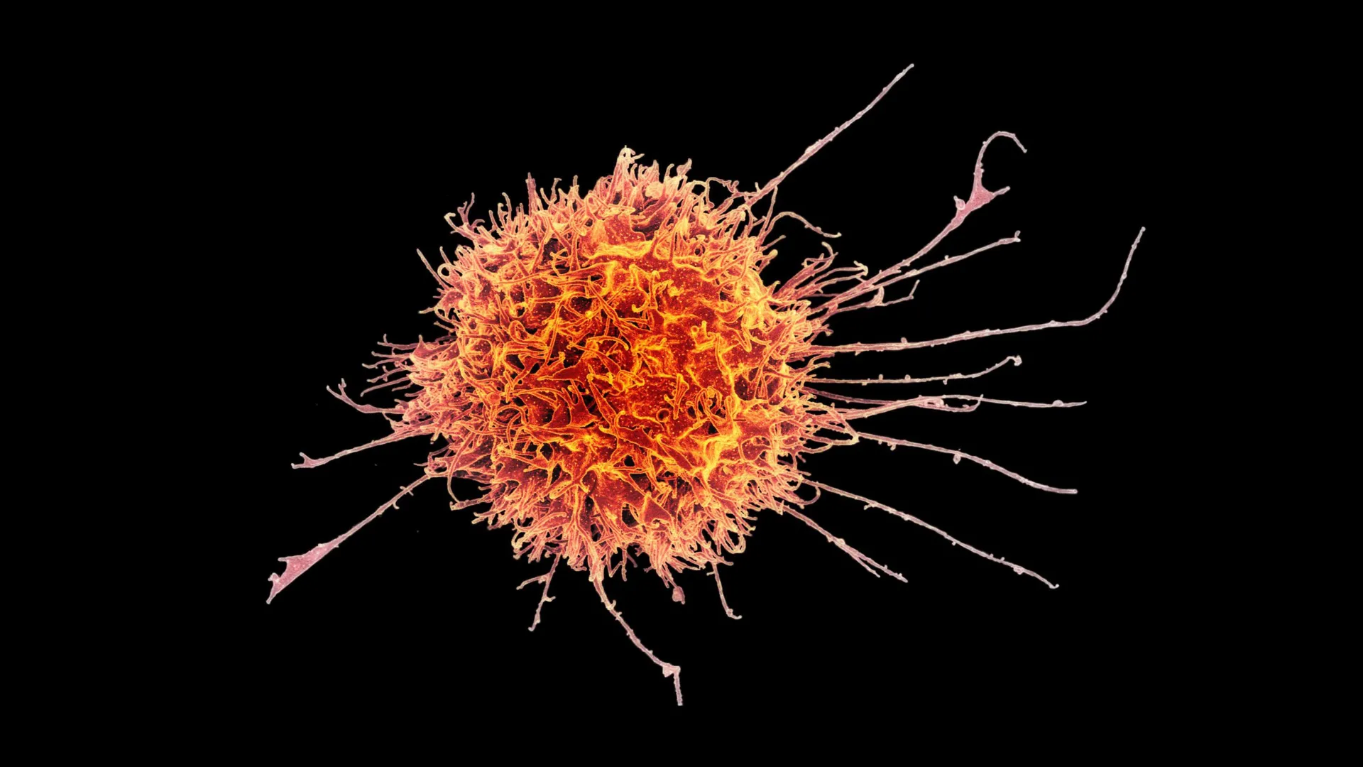
The landscape of oncology is currently undergoing a paradigm shift as researchers pivot from systemic treatments toward highly localized, cellular-level interventions. While chimeric antigen receptor (CAR) T-cell therapies have achieved remarkable success in treating hematologic malignancies, such as leukemia and lymphoma, the "solid tumor barrier" has remained a formidable challenge for the scientific community. Solid tumors, which account for approximately 90% of adult cancer cases, are notoriously difficult to treat because they reside within a hostile microenvironment that physically excludes immune cells and chemically suppresses their activity.
In a landmark study recently published in Science Translational Medicine, a multidisciplinary team led by researchers at Stanford Medicine has unveiled a novel strategy to breach these defenses. By reprogramming natural killer (NK) cells into a specialized "tissue-resident" state, the team has demonstrated a significantly enhanced ability to infiltrate solid tumors and execute targeted destruction of malignant cells. This development not only offers a potential new weapon against stubborn cancers like melanoma and head and neck squamous cell carcinoma but also paves the way for "off-the-shelf" cellular therapies that could be produced at scale and administered without the logistical hurdles of patient-specific cell manufacturing.
The Evolution of Immunotherapy and the Solid Tumor Challenge
To understand the significance of the Stanford breakthrough, one must look at the historical trajectory of cancer immunotherapy. For decades, the primary focus of immunology was centered on cells circulating in the peripheral blood. This "patrolling" immune system—consisting of B cells, T cells, and natural killer cells—was seen as the primary defense mechanism against systemic threats. However, recent advancements in bioinformatics and high-resolution imaging have revealed that the most critical immune interactions often occur within the tissues themselves.
Natural killer cells, first identified in the 1970s, are the body’s "first responders." Unlike T cells, which require prior exposure to a specific antigen to recognize a threat, NK cells possess an innate ability to detect and destroy cells that appear abnormal, whether due to viral infection or malignant transformation. Despite this inherent potency, conventional NK cells often fail when facing solid tumors. The tumor microenvironment (TME) acts as a fortress, utilizing dense extracellular matrices to block entry and secreting inhibitory signals like transforming growth factor-beta (TGF-β) to disarm any immune cells that manage to penetrate the perimeter.
"For a long time, the study of immunology and disease in humans was concentrated on the blood immune cells," noted John Sunwoo, MD, the Edward C. and Amy H. Sewall Professor in the School of Medicine and senior author of the study. "With the advancement of tools and bioinformatics, we are now starting to look more at what’s going on in tissue. For most immune cells, the tissue is where the action is."
The "Goldilocks" Discovery: Engineering Tissue Residency
The core of the Stanford team’s innovation lies in their ability to manipulate the developmental pathway of NK cells. While tissue-resident NK cells naturally exist in organs like the liver, lungs, and skin, they are notoriously difficult to harness for therapy because they can exist in two diametrically opposed states: one that is highly aggressive and one that is immunosuppressive.
Through a series of rigorous experiments, Sunwoo’s team discovered that the signaling protein TGF-β acts as a master regulator for these cells. However, its effect is highly dependent on the "dosage" and duration of exposure—a phenomenon the researchers describe as a "Goldilocks" effect.
The team found that prolonged exposure to TGF-β, which is common within the suppressive environment of a tumor, rendered NK cells dysfunctional and exhausted. Conversely, a carefully timed, high-intensity burst of TGF-β signaling, combined with direct physical contact with epithelial tumor cells, transformed circulating NK cells into a potent, tissue-resident form. These engineered cells expressed specific surface proteins—CD49a, CD103, and critically, CD39—that marked them as elite "killers" capable of surviving and thriving within the dense architecture of a solid tumor.
The researchers achieved this transformation by briefly exposing healthy donor NK cells to short-lived human epithelial tumor cells. This temporary interaction provided the necessary activating signals without the long-term exhaustion associated with chronic TGF-β exposure. The resulting cells were packed with perforin and granzyme A—the molecular tools used to puncture and dissolve cancer cells from the inside out.
Preclinical Success and Synergistic Potential
The efficacy of these modified tissue-resident NK (trNK) cells was tested in various mouse models of human cancer. The results were both reproducible and striking. When injected into mice bearing human melanoma and head and neck squamous cell carcinoma, the trNK cells demonstrated a superior ability to migrate into the heart of the tumors compared to standard, circulating NK cells.
The study also highlighted a powerful synergy between the modified cells and existing monoclonal antibody treatments. When paired with cetuximab—a drug already FDA-approved for certain colorectal and head and neck cancers—the trNK cells showed a dramatic increase in tumor suppression. Cetuximab works by binding to the epidermal growth factor receptor (EGFR) on cancer cells, effectively "flagging" them for immune destruction.
In the mouse models, a single dose of the combination therapy was able to halt tumor growth for over a month. While the control groups and those receiving monotherapies saw rapid disease progression, the mice receiving the combination treatment remained healthy and active through the 30-day observation period. "Even at day 30, when the other mice were sick, the mice that received the combination seemed very healthy," Sunwoo said, though he emphasized that these results are a "proof of concept" that must now be validated in human subjects.
A New Frontier: The "Off-the-Shelf" Advantage
One of the most significant implications of this research is its potential to democratize access to cell therapy. Currently, the most successful cell therapies, like CAR-T, are "autologous." This means they must be manufactured using a patient’s own cells—a process that is incredibly expensive (often costing hundreds of thousands of dollars), time-consuming (taking weeks to produce), and logistically complex.
Natural killer cells offer a distinct biological advantage: they generally do not cause graft-versus-host disease (GvHD). This means that NK cells from a healthy donor can be safely transferred to a patient without the need for perfect genetic matching.
Sunwoo’s team has already developed a scalable method for producing these trNK cells. According to their data, a single blood donation could yield enough cells to produce approximately 20 treatment doses within a two-week window. These doses can be cryopreserved, stored, and shipped to hospitals worldwide.
"It would be almost an off-the-shelf drug," Sunwoo explained. "It could make cell therapy much more accessible to a wider variety of patients." This scalability could address one of the primary criticisms of modern immunotherapy: that it is a "boutique" treatment available only to those at elite medical centers.
Chronology of the Research and Future Directions
The journey from the initial discovery of NK cells to this latest breakthrough spans half a century of immunological progress:
- 1970s: Identification of Natural Killer cells as a distinct lineage of lymphocytes.
- 1990s-2000s: Discovery of TGF-β as a potent immunosuppressor in the tumor microenvironment.
- 2010s: Success of CAR-T therapy in blood cancers highlights the potential of cell-based medicine.
- 2020-2023: The Stanford team identifies the specific signaling requirements (TGF-β and physical contact) to create cytotoxic tissue-resident NK cells.
- 2024: Publication of the findings in Science Translational Medicine and filing of patents for the production process.
Looking ahead, the research is moving rapidly toward clinical application. Sunwoo and his colleagues are in the process of preparing a Phase I clinical trial, which will evaluate the safety and preliminary efficacy of the combination therapy in patients with advanced squamous cell carcinoma. Pending approval from the U.S. Food and Drug Administration (FDA), the trial could begin as early as late 2024 or early 2025.
Broad Implications for Oncology and Immunology
The implications of this study extend beyond a single type of cancer. If NK cells can be reliably engineered to reside in tissues, this platform could potentially be adapted for any solid tumor that currently resists T-cell infiltration. Furthermore, the identification of CD39 as a marker for high-potency killer cells provides a new metric for researchers to evaluate the quality of immune cell products before they are administered to patients.
While the scientific community remains cautious—noting that mouse models do not always perfectly predict human responses—the Stanford study represents a significant leap forward in our understanding of how to "re-program" the innate immune system. By moving the focus from the bloodstream to the tissue, researchers are finally meeting cancer on its own turf.
As the medical community awaits the results of the upcoming Phase I trials, the Stanford breakthrough serves as a testament to the power of basic science. By deciphering the "Goldilocks" signaling of a single protein, researchers may have unlocked a way to transform the body’s first responders into its most effective assassins against solid tumors.
The research was a collaborative effort involving scientists from Ohio State University and Washington University School of Medicine. Funding was provided by the National Institutes of Health, the Tai Tsun Wu Research Fund for Natural Killer Cell Immunotherapy, and the Stanford Bio-X Fellowship.
