
 By Nana O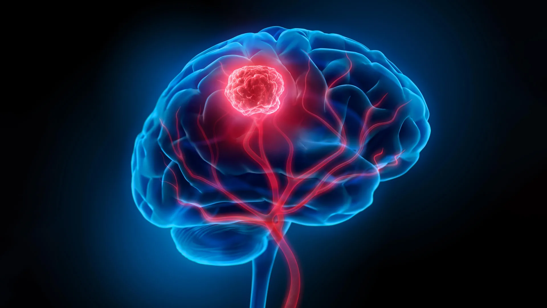
Glioblastoma, a formidable and highly aggressive form of brain cancer, has long presented a formidable challenge to medical science. Characterized by its rapid growth and invasive nature, glioblastoma tumors infiltrate surrounding healthy brain tissue, making complete surgical resection exceedingly difficult. Surgeons face a delicate balancing act, striving to remove cancerous cells while preserving vital neurological function. Compounding this challenge is the blood-brain barrier, a protective physiological shield that severely limits the efficacy of many therapeutic agents, including chemotherapy drugs and radiotherapy, in reaching tumor sites. These intertwined obstacles contribute to a grim prognosis, with the five-year survival rate for glioblastoma patients hovering at a mere 7 percent, underscoring the urgent need for innovative treatment strategies.
In a significant stride towards overcoming these persistent hurdles, researchers from the University of Technology Sydney (UTS), in collaboration with Harvard University and Henan University, have unveiled a groundbreaking ‘double-punch’ nanozyme platform. This sophisticated system, detailed in a recent publication in the esteemed journal Science Translational Medicine, leverages a single, intelligent nanoparticle design to address both the surgical and therapeutic limitations inherent in glioblastoma treatment.
A Synchronized Strategy: Imaging and Treatment in One
The core innovation of this new platform lies in its ability to perform two distinct yet complementary functions sequentially, all orchestrated by a single, engineered material. Dr. Bingyang Shi, Chair Professor of Nanomedicine at UTS’s School of Electrical, Mechanical and Biomedical Engineering, articulated the essence of this dual-action system: "We’ve engineered a single material that does two jobs in sequence. It’s a precise guide for the surgeon during the operation, and then a targeted clean-up treatment afterward."
At the heart of this platform is an exceptionally thin, two-dimensional sheet. This material is meticulously constructed atom by atom, employing techniques adapted from the advanced semiconductor manufacturing industry. This atomic-level precision in construction imbues the material with its remarkable versatility, enabling it to transition seamlessly between its imaging and therapeutic roles. Crucially, both of these functions are activated by the same specific wavelength of near-infrared light, a non-invasive energy source that can penetrate biological tissues.
Illuminating the Invisible: Enhanced Surgical Precision
The initial deployment of the nanozyme platform occurs during the surgical phase. Here, it acts as a highly sensitive imaging agent, significantly enhancing the surgeon’s ability to visualize the extent of the tumor. "During surgery, it functions as a highly sensitive imaging agent," explained Professor Shi. "A fluorescent dye engineered onto the sheet glows under a near-infrared wavelength invisible to the naked eye, allowing surgeons to see individual tumor cell clusters as small as 44 micrometers, a resolution beyond current clinical imaging tools." This level of detail is critical, as it surpasses the capabilities of many conventional intraoperative imaging modalities, enabling the detection of microscopic tumor deposits that might otherwise be missed.
Furthermore, the nanozyme platform is equipped with a targeting molecule. This crucial component is designed to facilitate its passage across the formidable blood-brain barrier and ensure its specific accumulation within glioma cells, thereby concentrating the imaging signal precisely where it is needed most. This targeted accumulation minimizes off-target effects and maximizes the clarity of the tumor margins visible to the surgical team.
Post-Operative Eradication: Targeting Residual Cancer Cells
Following the meticulous removal of the macroscopic tumor, the nanozyme platform is repurposed for a secondary, therapeutic role. After the visible tumor mass has been resected, the material is introduced into the surgical cavity. Upon re-activation with the same near-infrared light, the nanoparticles initiate a powerful anti-cancer effect.
"After the visible tumor is removed, the same material is administered into the surgical cavity and reactivated with the same wavelength of light for postoperative phototherapy," Professor Shi elaborated. The therapeutic mechanism is multifaceted. The platinum atoms embedded within the nanozyme structure play a pivotal role by catalyzing the conversion of the tumor’s own hydrogen peroxide into oxygen. This process is vital because glioblastoma tumors often create a hypoxic, or low-oxygen, microenvironment. This oxygen-depleted state typically shields cancer cells from conventional treatments. By increasing oxygen levels, the nanozyme platform disrupts this protective shield, making any remaining cancer cells more vulnerable.
Simultaneously, the near-infrared light activation triggers the generation of heat and reactive oxygen species. These highly energetic molecules are potent agents of cellular destruction, capable of eradicating microscopic cancer cells that surgery, by its very nature, cannot completely eliminate. This post-operative phototherapy is specifically designed to target and destroy any residual tumor cells lurking in the surgical bed, thereby addressing one of the most significant drivers of glioblastoma recurrence.
Addressing the Specter of Recurrence
The persistence of microscopic cancer cells after surgery is a central problem in glioblastoma management. Even when a surgeon believes they have achieved complete resection, these minuscule remnants can lie dormant before proliferating and initiating tumor regrowth. The dual-action nanozyme platform directly confronts this challenge. By providing enhanced visualization during surgery and then delivering a targeted therapeutic intervention to the surgical cavity, the technology aims to significantly reduce the likelihood of tumor recurrence.
Pre-Clinical Validation: Promising Results in Animal Models
The efficacy of this novel approach has been rigorously tested in pre-clinical studies using mouse models of glioblastoma. The results have been highly encouraging. In these studies, mice treated with the nanoparticle-enhanced surgical approach demonstrated a marked reduction in tumor recurrence following surgery. Notably, every mouse in the treated group remained alive at the 60-day mark, a significant improvement compared to the control group that received surgery alone, where the average survival was 42 days.
Beyond efficacy, the researchers also assessed the safety profile of the treatment. Follow-up examinations revealed no detectable neurological or motor impairments in the treated mice, suggesting a favorable safety margin for the nanozyme platform. This finding is crucial, as any new treatment for brain cancer must not only be effective but also preserve or minimize damage to delicate neural structures.
A Beacon of Hope, with Caveats
While the findings are exceptionally promising, the research team is keen to emphasize the early stage of this development. "The results are very encouraging, but this is still early-stage research carried out in mouse models, not in people – and that distinction is important," Professor Shi stressed. The transition from animal models to human clinical trials is a complex and lengthy process, involving extensive safety testing and efficacy evaluations.
The next critical steps involve scaling up the technology to be applicable to the human brain, which is significantly larger and more complex than a mouse brain. "Its imaging and therapeutic performance will also need to be confirmed at the scale of a human brain," Professor Shi noted. This will require further refinement of the nanoparticle design, manufacturing processes, and delivery mechanisms to ensure consistent and effective performance in a human patient.
Broader Implications for Neuro-Oncology
Should this technology successfully navigate the rigorous pathway of clinical trials and gain regulatory approval, it holds the potential to fundamentally alter the landscape of glioblastoma treatment. For patients, it could mean a more precise and less invasive surgical experience, with surgeons empowered to identify and excise more of the tumor. The subsequent targeted therapy offers a crucial safeguard against the insidious threat of recurrence, a prospect that weighs heavily on the minds of glioblastoma patients and their families.
The development represents a significant advancement in the field of nanomedicine and its application to oncology. By creating a single platform that integrates diagnostic and therapeutic capabilities, the researchers have exemplified a sophisticated approach to tackling complex diseases. This ‘theranostic’ strategy – combining therapy and diagnostics – is gaining increasing traction in medical research due to its potential for personalized and more effective patient care.
The potential implications extend beyond glioblastoma. The principles behind this dual-function nanozyme platform—its ability to cross the blood-brain barrier, its targeted accumulation, and its light-activated dual action—could theoretically be adapted to target other neurological diseases or brain tumors with similar invasive characteristics. This adaptability underscores the broader scientific value of the discovery.
Professor Shi’s concluding remarks encapsulate the forward-looking optimism: "If this continues to hold up through that process, the hope is that surgeons could one day see more of the tumor during an operation and treat more of what’s left behind afterward. It’s a meaningful step towards reducing recurrence, which remains one of the biggest challenges for people with glioblastoma." This research offers a tangible glimpse into a future where advanced nanotechnology plays a pivotal role in conquering some of the most challenging diseases known to humankind. The journey from laboratory discovery to patient bedside is arduous, but this innovative nanoparticle platform represents a compelling and hopeful stride forward.
