
 By Budi Oetomo Narendra
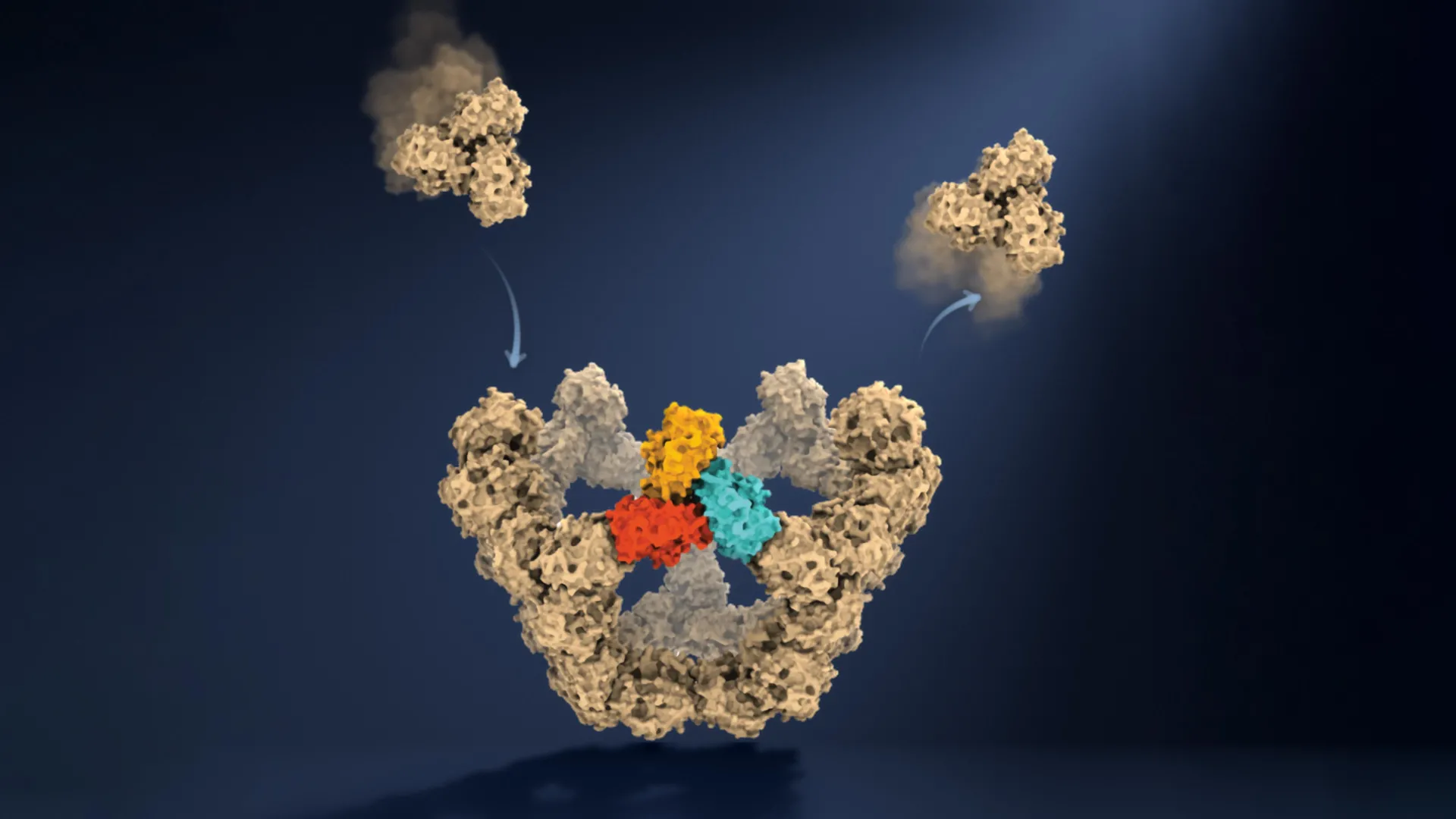
Researchers at the University of Oxford have achieved a significant breakthrough, observing in real-time how virus-like particles meticulously assemble themselves, molecule by molecule. This unprecedented insight, detailed in a new study published in the prestigious journal Nature, reveals the surprisingly simple yet remarkably reliable physical rules governing the formation of complex biological structures. For decades, scientists have grappled with directly observing this fundamental process, making this discovery a pivotal moment in virology and molecular biology. The findings not only shed light on a long-standing scientific enigma but also open new avenues for developing advanced antiviral drugs, more effective vaccines, and a host of other medical treatments.
A Decades-Long Scientific Enigma: The Challenge of Viral Construction
Viruses, despite their microscopic size and apparent simplicity, present a profound biological paradox. They are essentially genetic material encased within a protective protein shell, known as a capsid. These capsids, which can comprise anywhere from tens to thousands of individual protein subunits, must assemble with astounding precision to form robust, functional structures capable of shielding their precious genetic cargo and facilitating infection. What has long puzzled scientists is how these individual protein components consistently self-organize into the correct, highly symmetrical structures – often icosahedral or helical – without any external blueprint or complex cellular machinery directing the process. This spontaneous, self-correcting assembly, occurring within milliseconds, represents an extraordinary feat of molecular engineering, a problem that nature has elegantly solved.
For much of the 20th century, understanding viral assembly relied heavily on theoretical models and static images obtained through techniques like electron microscopy. Pioneering work, such as the Caspar-Klug theory developed in the late 1950s and early 1960s, provided mathematical frameworks to describe the geometry of viral capsids, suggesting how identical protein subunits could arrange into larger, stable structures. However, these models, while foundational, could only infer the dynamic process of assembly. Experimental observations were largely limited to examining the starting materials and the finished viral particles. The fleeting intermediate structures, which exist for mere moments and in minuscule quantities, remained elusive, making it impossible to truly understand the kinetic pathways and error-correction mechanisms at play. This gap in direct observation represented a significant bottleneck in developing targeted interventions against viral replication. The challenge was akin to trying to understand how a skyscraper is built by only seeing the raw materials and the finished building, without ever observing the construction crew or the stages of erection.
A New Window into Molecular Dynamics: The Mass Photometry Breakthrough
To overcome these formidable observational limitations, the Oxford researchers employed an innovative methodology centered around mass photometry, a cutting-edge technique developed at the University of Oxford. Mass photometry operates on a remarkably simple yet powerful principle: it determines the mass of individual molecules by precisely measuring the amount of light they scatter when illuminated. Unlike traditional techniques that average signals from millions of molecules, mass photometry provides single-molecule resolution, making it uniquely suited for studying transient biological processes.
For this study, the team focused on an engineered virus-like particle (VLP) composed of 60 protein units. VLPs are non-infectious protein shells that mimic the structure of actual viruses but lack genetic material, making them safe and controllable models for studying assembly. The choice of a relatively simple, well-characterized VLP allowed the researchers to establish a clear baseline for observation. The true ingenuity of their approach, however, lay in combining mass photometry with a novel method for confining individual molecules and continuously observing them over extended periods. This allowed the scientists to repeatedly measure the mass of a single, growing particle as additional protein components attached to it.
"This method lets us study the assembly process directly, we just take a video!" explained co-first author PhD student Dan Loewenthal from the Department of Chemistry and Kavli Institute for Nanoscience Discovery at the University of Oxford. This analogy perfectly encapsulates the transformative nature of their technique. Instead of capturing static "snapshots" at different points, they were able to record the continuous addition of individual molecular building blocks, effectively producing a real-time molecular movie of capsid construction. This capability represents a paradigm shift, moving from inferring mechanisms based on endpoints to directly observing the kinetics and thermodynamics of assembly pathways.
Professor Philipp Kukura, also a co-lead author from the Department of Chemistry and Kavli Institute for Nanoscience Discovery, emphasized the significance: "Until now, much of our understanding of how these structures assemble has had to be reconstructed from snapshots or theoretical models. Being able to both quantify the underlying interactions and follow one particle as it grows changes that. We can see the important intermediate structures appear and deduce a detailed model of the construction process." This direct observation provides empirical data to validate or refine existing theoretical models and offers unprecedented detail into the nuances of molecular interactions.
Unveiling Nature’s Design Principles: The Mechanism of Reliable Assembly
The real-time observation unveiled a fascinating and highly efficient mechanism for reliable self-assembly. The researchers discovered that viral assembly is not a chaotic process of random collisions but rather resembles "navigating a complicated maze," where only specific pathways lead to lasting progress and the formation of the correct structure. The key to this efficiency lies in a sophisticated interplay of weak and strong molecular interactions, coupled with an inherent error-correction mechanism.
Initially, protein components form weak, transient connections. These initial bonds are relatively unstable and can easily break apart if the arrangement is incorrect or unfavorable. This inherent flexibility is crucial; it allows the molecules to "try out" different combinations, making mistakes without permanent consequence. If an arrangement is unsuccessful, the weakly bound components can dissociate, giving them another opportunity to find a better, more stable configuration. This trial-and-error approach prevents the system from getting stuck in kinetically trapped, non-functional structures.
Once multiple components eventually connect in a particular closed structure, the collective strength of these multiple bonds renders the arrangement far more stable. These stable arrangements act as "molecular checkpoints," effectively narrowing the vast number of theoretically possible assembly routes into a much smaller, manageable set of successful pathways. The most critical of these milestones, the researchers found, occurs when five larger protein building blocks connect to form a closed pentagonal ring. This pentagonal ring represents the first highly stable structure in the entire assembly process. Its formation is a bottleneck, but once achieved, it provides a robust platform for subsequent growth.
Dr. Roi Asor, a co-lead author from the Department of Chemistry and Kavli Institute for Nanoscience Discovery, articulated the challenge and the solution: "A virus has to solve an extraordinary construction problem. Its components somehow have to find the right arrangement among a huge number of possibilities, without a blueprint or machinery directing the process. We can now watch what happens molecule by molecule and see the physical rules that make it possible." He further elaborated on the elegance of the system: "The weak interactions give the system room to make mistakes. Most encounters don’t have to be successful: the components can separate and try again. But once enough of them come together in the right closed arrangement, the structure becomes stable, and assembly can move forward. That combination of trial and error followed by locking in successful structures is what makes the process so reliable."
The findings also clarify why the earliest stage of construction, known as nucleation, is particularly challenging and often the slowest step. Nucleation requires a critical mass of correctly oriented components to come together and form the first stable intermediate. Once this initial stable structure (like the pentagonal ring) forms, each subsequent stable stage requires fewer additional building blocks, and the growth rate accelerates significantly. This explains the characteristic S-shaped kinetics observed in many self-assembly processes, where there is an initial slow lag phase followed by rapid growth.
Voices from the Forefront: Researcher Perspectives
The research team expressed profound excitement regarding the implications of their discovery. Their collective sentiments underscored both the scientific elegance of the findings and their practical potential.
Dan Loewenthal’s simple analogy of "taking a video" highlights the directness and clarity of their observational method, a stark contrast to previous indirect approaches. This directness offers a level of certainty and detail previously unattainable.
Professor Philipp Kukura emphasized the shift from theoretical reconstruction to empirical quantification. His statement about "being able to both quantify the underlying interactions and follow one particle as it grows" points to a new era of understanding the precise energetic and kinetic parameters that govern self-assembly, moving beyond mere descriptive models. This quantitative understanding is crucial for predictive science and engineering.
Dr. Roi Asor’s explanation eloquently captured the essence of the viral construction problem and how the observed physical rules provide an answer. His focus on the "combination of trial and error followed by locking in successful structures" distills the core principle of reliable self-assembly, a principle that may extend far beyond viruses.
Dr. Jack Tan, a co-author from the MRC Weatherall Institute of Molecular Medicine and CAMS-Oxford Institute, specifically highlighted the translational potential, noting that "The ability to understand these processes at the molecular level could have important applications in vaccine development and antivirals that disrupt viral assembly." His perspective bridges the gap between fundamental research and practical medical applications, underscoring the immediate relevance of the findings to public health. The collective enthusiasm from the Oxford team resonates with the broader scientific community, which has long sought such detailed insights into fundamental biological processes.
Revolutionizing Medicine: Implications for Antivirals and Vaccines
The molecular-level understanding of viral assembly gained from this study holds immense promise for revolutionizing the development of antiviral drugs and vaccines. Current antiviral strategies often target viral enzymes or inhibit replication at later stages of the viral life cycle. However, directly interfering with the construction of the protective capsid offers a new and potentially highly effective therapeutic avenue.
By identifying the specific molecular interactions that control the stability of intermediate structures and the transition points (molecular checkpoints) in the assembly process, scientists could design drugs that specifically target these vulnerabilities. For instance, a drug could be engineered to disrupt the weak initial connections, preventing stable nucleation altogether. Alternatively, an antiviral could bind to and destabilize crucial intermediate structures, such as the pentagonal ring, thereby preventing the particle from reaching completion. This "assembly interference" strategy could be particularly potent because it targets a fundamental and often conserved aspect of viral biology. Many viruses, despite their diversity, share similar architectural principles for capsid formation.
Moreover, the insights could significantly advance vaccine development. Virus-like particles (VLPs) are already widely used as vaccine platforms, notably in the human papillomavirus (HPV) vaccine. VLPs are highly immunogenic because their structural proteins mimic the surface antigens of actual viruses, prompting a robust immune response without the risk of infection. A deeper understanding of self-assembly principles would enable scientists to engineer more stable, more efficiently produced, and potentially more immunogenic VLPs. For example, by optimizing the interactions between protein subunits, researchers could design VLPs that assemble more reliably in industrial quantities, or even design them to incorporate specific antigens more effectively for broader protection against different viral strains. This could lead to next-generation VLP vaccines that are more potent, safer, and easier to manufacture, addressing global health challenges more effectively.
Beyond the Viral Realm: Broader Biological and Nanotechnological Impact
The implications of this research extend far beyond the immediate context of viruses. Self-assembly is a universal principle fundamental to countless biological processes, orchestrating the formation of virtually every complex structure within living organisms. From the intricate internal skeletons of cells (cytoskeleton) and the machinery that translates genetic code (ribosomes) to specialized molecular compartments and large protein complexes crucial for cellular function, self-assembly is the underlying mechanism.
This new methodology, allowing for the observation of intricate construction processes one molecule at a time, offers researchers a powerful new tool to investigate how these complex biological structures emerge from relatively simple molecular interactions. For example, understanding the assembly pathways of amyloid fibrils, misfolded protein aggregates implicated in neurodegenerative diseases like Alzheimer’s and Parkinson’s, could lead to novel therapeutic strategies. By identifying the critical nucleation steps or stable intermediates in amyloid formation, interventions could be designed to prevent or reverse their pathological accumulation.
Furthermore, the principles elucidated by the Oxford team hold significant promise for the burgeoning fields of nanoscience and materials engineering. Scientists and engineers are increasingly seeking to design novel materials that can self-assemble into complex, functional architectures from simple building blocks. Nature has perfected this art over billions of years, and by deciphering its rules, researchers can draw inspiration to create advanced nanomaterials for applications ranging from drug delivery systems and biosensors to catalysts and energy storage devices. The ability to control self-assembly at the molecular level could unlock new frontiers in creating bespoke materials with unprecedented precision and functionality.
In conclusion, the Oxford team’s ability to "take a video" of viral assembly represents a monumental leap in our understanding of how complex biological structures are formed. By revealing the elegant dance of trial-and-error, weak interactions, and molecular checkpoints, this research not only solves a decades-old scientific mystery but also lays critical groundwork for developing innovative medical therapies and inspiring a new generation of self-assembling materials. It underscores the profound truth that sometimes, the most complex problems are solved by surprisingly simple, yet robust, physical rules.
