
 By Gabriel Paijo
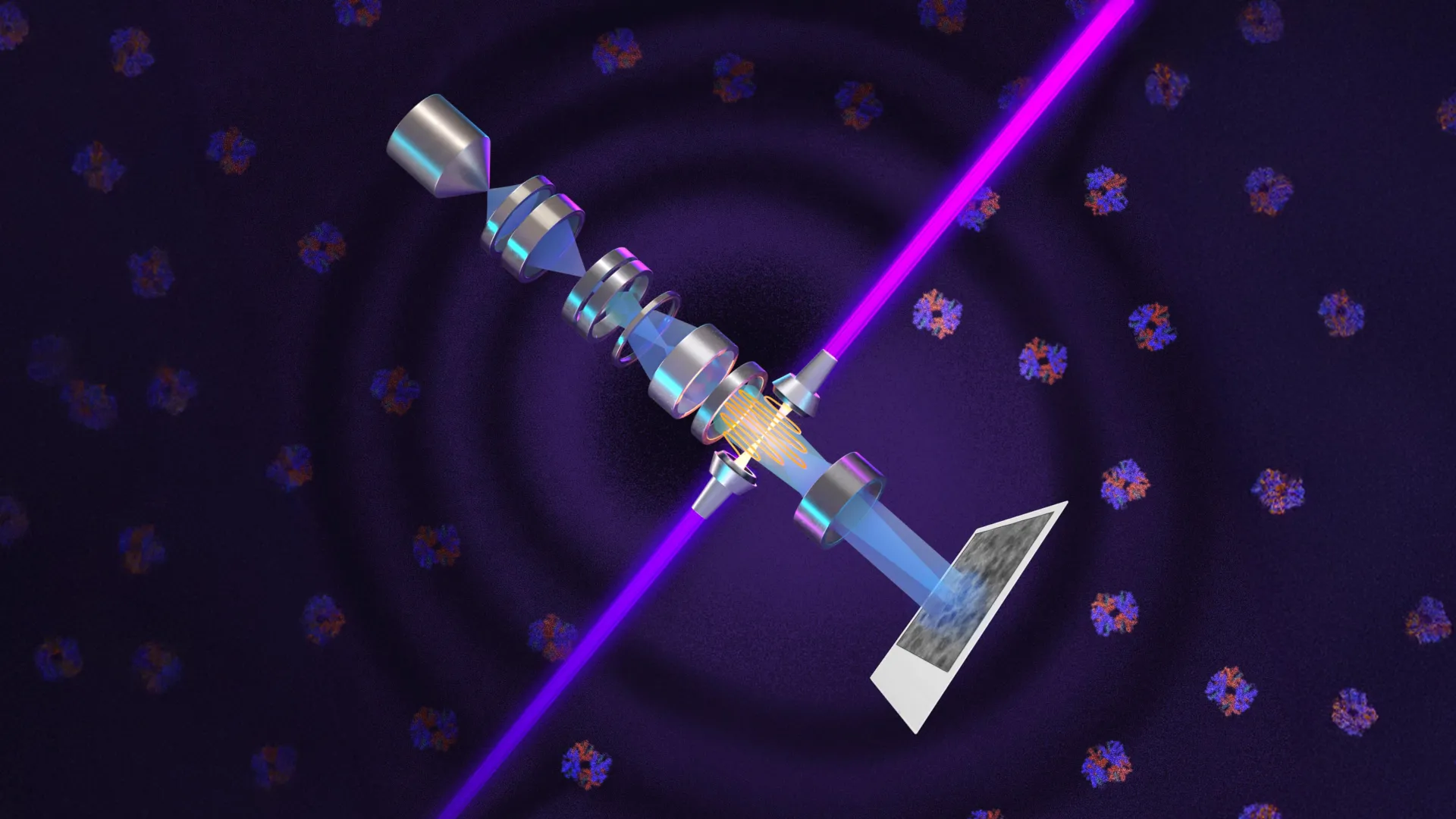
A team of physicists at the University of California, Berkeley, has successfully integrated a Nobel Prize-winning optical principle with state-of-the-art electron microscopy, creating a technological leap that allows scientists to visualize the smallest building blocks of life with unprecedented clarity. By developing a device known as a laser phase plate, the researchers have addressed a century-old limitation in biological imaging, potentially opening a window into the "invisible" 90% of the human proteome. This breakthrough, led by UC Berkeley professor Holger Müller and faculty scientist at Lawrence Berkeley National Laboratory, utilizes an extraordinarily intense continuous-wave laser to enhance the contrast of biological specimens, enabling the study of proteins that were previously too small or too faint to be analyzed.
The development, detailed in the journal Science, marks the culmination of 15 years of engineering and theoretical research. The new technology is primarily aimed at improving cryoelectron microscopy (cryo-EM) and cryoelectron tomography (cryo-ET), two methods that have revolutionized structural biology by allowing researchers to see molecules in their near-native states. Despite their success, these methods have long struggled with a fundamental "signal-to-noise" problem: most human proteins are so small that they scatter very few electrons, making them appear nearly transparent against their background. The laser phase plate solves this by shifting the phase of the electron beam, effectively turning invisible structures into high-contrast images without damaging the delicate biological samples.
The Evolution of Phase Contrast: From Light to Electrons
The journey toward this discovery began nearly a century ago. In the early 1930s, Dutch physicist Frits Zernike addressed a persistent problem in light microscopy. Because biological cells are composed mostly of water, they are almost entirely transparent. While researchers could use chemical stains to create contrast, the process typically killed the cells, preventing the study of live biological processes. Zernike realized that while light passing through a cell might not change much in brightness, it does experience a tiny delay, or phase shift, as it interacts with cellular structures.
Zernike’s innovation was to develop a "phase plate" that could shift the timing of unscattered light waves by 90 degrees. When these waves recombined with light scattered by the sample, the interference created visible differences in brightness. This technique, known as phase-contrast microscopy, earned Zernike the Nobel Prize in Physics in 1953 and became a staple of biological research. However, applying this principle to electron microscopy—which uses electron beams instead of light to achieve 10,000 times higher magnification—proved to be an immense technical challenge.
Early attempts to create phase plates for electron microscopes involved placing thin physical films in the path of the electron beam. These films were problematic; they often became contaminated, weakened the beam, or introduced optical distortions that blurred the resulting images. The quest for a "clean" phase shift remained unfulfilled for decades until Müller and UC Berkeley professor emeritus Robert Glaeser proposed a radical solution in 2010: using a high-intensity laser instead of a physical material to alter the electron beam’s phase.
Engineering the World’s Most Intense Continuous Laser Focus
To achieve a 90-degree phase shift in an electron beam, the UC Berkeley team needed to generate a laser field of staggering intensity. The solution involved a complex optical system that confines a continuous laser beam inside a spherical chamber lined with ultra-reflective mirrors. As the light bounces between these mirrors more than 10,000 times, the energy builds up, creating a focal point of 75 kilowatts concentrated into a spot only a few microns wide.
"It’s more powerful than what you use for welding. It’s more power than a military laser," Müller noted regarding the intensity of the focus. This "laser phase plate" interacts with the electrons passing through it via the ponderomotive force, a non-linear force that pushes electrons toward areas of lower optical intensity. By precisely positioning this laser focus, the researchers can delay the phase of the electron beam just enough to create the interference patterns necessary for high-contrast imaging.
The team integrated this technology into a customized Thermo Fisher Krios cryo-EM instrument, which they named "Theia" after the Greek Titaness of light. Theia is described by the researchers as a "Formula 1" microscope, featuring advanced electron optics that provide superior resolution even before the laser is activated. With the addition of the laser phase plate, the instrument is positioned to become the most capable biological imaging tool in existence.
Overcoming the 70-Kilodalton Barrier
The primary metric for measuring protein size is the dalton (Da), with one dalton roughly equal to the mass of a single hydrogen atom. In the world of structural biology, size is a major obstacle. Conventional cryo-EM techniques typically struggle to resolve proteins smaller than 70 kilodaltons (kDa). This is a significant limitation because the vast majority of proteins produced by the human body—estimated at roughly 90% of the proteome—fall below this size threshold.
In their validation tests, the Berkeley team used Theia to image two distinct proteins: aldolase and hemoglobin. While aldolase is a relatively large protein that is easily imaged by standard cryo-EM, hemoglobin is much smaller (approximately 64 kDa) and sits at the edge of what current technology can reliably see. The results were definitive: the laser phase plate significantly improved the resolution and contrast for both, but the improvement for hemoglobin was particularly striking.
The ability to resolve molecules at the 50 kDa level, and eventually down to 17 kDa (the size of myoglobin), would represent a "step function change" for biology. It would allow scientists to map the structures of thousands of proteins that have never been seen before because they are too small for cryo-EM and too difficult to crystallize for X-ray crystallography.
Chronology of Structural Biology Milestones
The development of the laser phase plate is the latest entry in a timeline of breakthroughs that have defined modern biochemistry:
- 1930s: Frits Zernike invents phase-contrast light microscopy.
- 1953: Zernike receives the Nobel Prize in Physics.
- 1960s-70s: Robert Glaeser and colleagues demonstrate that freezing biological samples (cryopreservation) can protect them from the radiation damage caused by electron beams.
- 2010: Holger Müller and Robert Glaeser publish the theoretical framework for a laser-based phase plate.
- 2017: Jacques Dubochet, Joachim Frank, and Richard Henderson receive the Nobel Prize in Chemistry for developing cryo-EM, acknowledging Glaeser’s foundational work.
- 2024: The UC Berkeley team publishes the first successful high-resolution images obtained using a continuous-wave laser phase plate in Science.
Collaborative Innovation and the Two-Laser Design
The success of the project was bolstered by a collaboration with the Chan Zuckerberg Biohub (CZ Biohub). While Müller’s team at Berkeley focused on the single-laser "Theia" system, researchers at the Biohub in Redwood City, led by Bridget Carragher and David Agard, are developing a parallel system using a two-laser configuration.
This dual-laser design, based on Müller’s theoretical research, arranges two beams at right angles. By splitting the power between two lasers, the system reduces the thermal load on individual components, preventing burnout and minimizing optical distortions. This approach is intended to make the technology more robust and easier to implement in commercial settings. Both teams are working closely with Thermo Fisher Scientific, the industry leader in electron microscope manufacturing, to explore how this technology can be integrated into the next generation of scientific instruments.
Implications for Drug Discovery and Disease Research
The impact of this technology extends far beyond the walls of physics laboratories. By improving the signal-to-noise ratio in cryo-EM, the laser phase plate will allow drug researchers to see how small-molecule drugs bind to their target proteins with much greater precision. Currently, many potential drug targets are "undruggable" simply because their structures cannot be resolved clearly enough to design effective interventions.
Furthermore, the technology is expected to be "revolutionary" for cryoelectron tomography (cryo-ET). Unlike standard cryo-EM, which looks at purified proteins in a solution, cryo-ET captures images of molecules inside their natural cellular environment. This is often compared to looking for a specific leaf in a dense forest.
"Cryo-ET needs a dramatic step forward in contrast so we can start to see what’s going on inside the cell," said Bridget Carragher. "That’s what the laser phase plate promises to give us."
By providing high-contrast, three-dimensional views of the "crowded" interior of a cell, researchers can observe how molecular machines operate in context. This could lead to breakthroughs in understanding how viruses infect cells, how proteins misfold in neurodegenerative diseases like Alzheimer’s, and how cancer cells evade the immune system.
Future Horizons: Reaching the 17-Kilodalton Goal
Looking ahead, Holger Müller and his team are already planning further refinements to the system. The next major goal is to enable the imaging of proteins as small as 17 kilodaltons. Achieving this may involve transitioning from the current "defocused" beam method to a "focused" electron beam approach.
Under current cryo-EM protocols, researchers often have to slightly defocus the microscope to generate enough contrast to see the molecules. However, this defocusing inherently limits the resolution. Müller believes the laser phase plate will eventually allow for "in-focus" imaging, which could potentially double the contrast and signal-to-noise ratio once again.
As Stephani Otte, Vice President of Imaging Science at CZ Biohub, noted, the ability to see what was once invisible changes the fundamental landscape of biological science. By bridging the gap between the physics of light and the mechanics of electrons, the UC Berkeley team has provided a new set of eyes for the scientific community, ensuring that the next century of cellular exploration will be as transformative as the last.
