
 By George Ongkojoyo
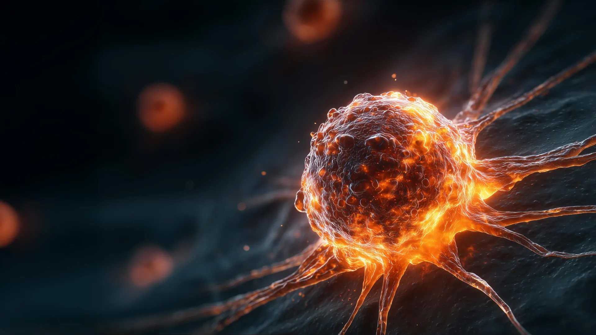
The complexity of cancer lies in its diversity; while some tumors remain localized and treatable, others exhibit a lethal propensity for rapid growth, aggressive metastasis, and profound resistance to conventional therapies. At the Fralin Life Sciences Institute at Virginia Tech, a multidisciplinary team of researchers has spent the last half-decade peeling back the layers of this biological enigma. Their findings, recently published in the Proceedings of the National Academy of Sciences (PNAS) and Cancer Research, reveal two previously misunderstood drivers of malignancy: the ability of abnormal cancer cells to manipulate healthy tissue into supporting tumor expansion and a counterintuitive link between the physical size of cancer cells and their level of danger to the patient.
These discoveries are the culmination of five years of rigorous experimentation, involving advanced genomic analysis, mouse model studies, and the meticulous microscopic examination of tumor architecture. By focusing on tetraploidy—a state where cells possess double the normal amount of genetic material—the researchers have identified a specific cellular "recruitment" strategy that allows tumors to thrive even when the most abnormal cells are in the minority. Furthermore, their identification of cell size as a prognostic marker offers a potential new tool for clinicians to predict patient outcomes more accurately.
The Microscopic Labor of Cancer Research
The path to these scientific breakthroughs begins in the quiet, climate-controlled laboratories of Virginia Tech, where graduate students like Megan Sweet perform the exacting work of histology. To understand how a tumor functions as a living organ, researchers must be able to see its internal structure at a cellular level. This requires the preparation of tissue samples so thin that they are nearly transparent.
Working within a specialized refrigerated metal chamber known as a cryostat, Sweet processes tumor samples roughly the size of a pinkie fingernail. The process is one of extreme precision; the tissue must be mounted at a perfect angle before a razor-sharp blade slices it into sections measured in microns. As the blade moves, the rhythmic sound of the machine underscores the repetitive nature of the work. For Sweet, this "meditative" process is the essential foundation of the study. Each slice is transferred to a glass slide and treated with specific chemical stains that highlight various cellular components—nuclei, membranes, and the surrounding connective tissue.
This painstaking methodology allowed the team to observe not just the cancer cells themselves, but how those cells interact with their environment. By comparing thousands of these stained slides, the team began to see a pattern: the most aggressive tumors were not necessarily those composed entirely of abnormal cells, but those that had most effectively "hired" the body’s own healthy cells to work on their behalf.
Chromosomal Instability and the Rise of Tetraploidy
To understand the team’s findings, one must first look at the fundamental blueprint of human life: the chromosome. Most healthy human cells are diploid, meaning they carry two complete sets of chromosomes—one from each parent. During normal cell division (mitosis), these chromosomes are replicated and then evenly distributed between two new daughter cells. However, this process is prone to error.
When a cell replicates its DNA but fails to complete the final stage of division, it results in a tetraploid cell—a cell containing four sets of chromosomes. While the body often identifies and eliminates these abnormal cells through programmed cell death (apoptosis), some survive. These survivors often possess a high degree of chromosomal instability, meaning they continue to gain or lose genetic material at an accelerated rate. This instability is a hallmark of many of the most difficult-to-treat cancers.
Under the guidance of cell biologist Daniela Cimini, graduate students Mat Bloomfield and Megan Sweet focused their research on these tetraploid cells. By using laboratory techniques to intentionally induce tetraploidy in human cancer cell lines, the team created a controlled environment to study how these cells behave compared to their "normal" diploid counterparts. What they found challenged long-standing assumptions about tumor composition and growth.
The Trojan Horse Effect: Recruiting the Stroma
One of the most significant findings of the Virginia Tech study involves the relationship between tetraploid cells and the tumor microenvironment. Scientists have known for decades that tetraploidy is associated with more aggressive cancers, but the prevailing theory was that these cells simply out-competed other cells.
The Virginia Tech team’s experiments in mouse models revealed a more complex reality. They observed that as a tumor grew, the actual percentage of tetraploid cells within the mass often decreased over time. Paradoxically, the tumors continued to grow at an accelerated rate. Upon closer inspection of the tissue slides, the researchers discovered that the tetraploid cells were acting as "recruiters."
These abnormal cells were found to be highly effective at attracting stromal cells—non-cancerous connective tissue cells that normally provide structural support and wound healing for healthy organs. In the context of a tumor, these recruited stromal cells are "hijacked" to provide a supportive scaffolding and growth signals that fuel the expansion of the remaining cancer cells.
"The presence of even a small fraction of these tetraploid cells can promote the recruitment of extra non-cancerous cells that support further tumor progression," Megan Sweet explained. This discovery suggests that even if a treatment successfully kills off the majority of highly abnormal tetraploid cells, the "damage" may already be done; the recruited healthy cells continue to provide an environment where the remaining cancer can flourish.
The Size Paradox: Why Smaller Cells Pose a Greater Risk
While investigating the physical properties of tetraploid cells, Mat Bloomfield made an observation that initially seemed like an error. Because tetraploid cells contain twice the DNA of normal cells, they are typically much larger in volume. However, as Bloomfield established colonies from individual tetraploid cells, he noticed a significant variance in size.
Some of these tetraploid colonies were 25 to 30 percent smaller than expected. Logically, one might assume that a larger, more genetically "bloated" cell would be more dangerous. The data proved the opposite. The smaller tetraploid clones were significantly more aggressive than the larger ones. These smaller cells demonstrated faster replication cycles, a higher capacity for invasion into surrounding tissues, and a notable resistance to common chemotherapy drugs and cellular stress.
This "size paradox" was consistent across different types of cancer, including colorectal and breast cancer cell lines. The researchers hypothesize that these smaller cells may have undergone a rapid "evolutionary" adjustment, shedding unnecessary cellular bulk while retaining the aggressive genetic advantages of their tetraploid origins. This streamlined state appears to make them more metabolically efficient and harder to kill.
Validating Findings through the Cancer Genome Atlas
To ensure their laboratory findings were relevant to human health, the Virginia Tech team turned to the Cancer Genome Atlas (TCGA), a landmark project co-managed by the National Cancer Institute and the National Human Genome Research Institute. The TCGA contains genomic and clinical data from over 20,000 primary cancer and matched normal samples spanning 33 cancer types.
By analyzing this massive data set, the researchers were able to correlate cell size and chromosome count with actual patient outcomes. The data confirmed their lab observations: patients whose tumors contained smaller tetraploid cells had significantly lower survival rates and more rapid disease progression than those with larger tetraploid cells.
This validation provides a bridge between basic laboratory science and clinical application. "We already knew that tetraploidy can make cells more tumorigenic," said Daniela Cimini. "But now we know that if you incorporate the size of the cells, it can be more predictive of tumorigenic potential."
Timeline of Discovery and Scientific Context
The Virginia Tech research adds a critical chapter to a century-old scientific narrative. The link between abnormal chromosome numbers and cancer was first proposed by German biologist Theodor Boveri in 1914. However, for much of the 20th century, the technology did not exist to observe these processes in real-time or to manipulate them with precision.
- 2019-2020: The Virginia Tech team begins isolating tetraploid cells to study their long-term viability.
- 2021-2022: Experiments in mouse models reveal the unexpected recruitment of stromal cells, shifting the focus from the cancer cells alone to the "neighborhood" they inhabit.
- 2023: Mat Bloomfield identifies the correlation between smaller cell size and increased drug resistance.
- 2024: The team publishes two major papers in Cancer Research and PNAS, solidifying the link between cell morphology and clinical prognosis.
Implications for Future Treatment and Diagnostics
The implications of this research for the future of oncology are twofold. First, it suggests a new diagnostic metric. Currently, pathologists examine tumor biopsies for "atypia"—general abnormalities in cell shape and size. By specifically measuring the volume of tetraploid cells, doctors may soon be able to assign a more precise "aggression score" to a patient’s cancer, allowing for more personalized treatment plans.
Second, the discovery of stromal recruitment opens the door for new therapeutic targets. If researchers can identify the specific chemical signals that tetraploid cells use to "call in" healthy tissue, they may be able to develop drugs that block those signals. By preventing the tumor from building its support network, the cancer could be made more vulnerable to existing treatments.
The work of Sweet, Bloomfield, and Cimini underscores the importance of fundamental biological research. By spending thousands of hours at the lab bench, slicing tissue and analyzing data, they have moved the scientific community one step closer to understanding the "how" and "why" behind the most dangerous forms of cancer. As the team continues to investigate the specific biological mechanisms that allow smaller cells to resist drugs, their work remains a testament to the power of meticulous, persistent inquiry in the fight against disease.
