
 By Gabriel Paijo
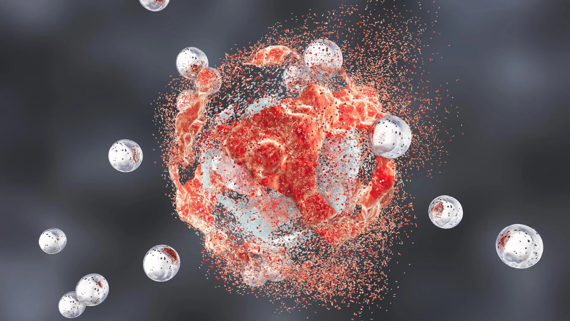
In a significant advancement for the field of nanomedicine, a multidisciplinary team of researchers from Weill Cornell Medicine and the Cornell Duffield College of Engineering has developed a novel therapeutic approach using engineered silica nanoparticles to combat aggressive prostate cancer. This preclinical study, recently published in the journal Cancer Research, details the development of "Cornell Prime dots" (C’ dots)—ultrasmall, fluorescent, core-shell silica particles that possess the unique ability to directly destroy tumor cells while simultaneously activating the body’s immune system. By leveraging a specialized form of cell death known as ferroptosis and transforming the immunological landscape of the tumor, these nanoparticles have achieved complete remissions in mouse models, signaling a potential shift in how refractory cancers are treated in a clinical setting.
The Evolution of C’ Dots: From Imaging to Therapy
The journey of these nanoparticles began not as a primary treatment, but as a sophisticated tool for medical imaging. Originally conceptualized by Dr. Ulrich Wiesner and his team at Cornell University’s Department of Materials Science and Engineering, the C’ dots were designed to be "ultrasmall"—typically less than 10 nanometers in diameter. Their size is a critical factor in their biological utility, as it allows them to circulate through the bloodstream, penetrate deeply into dense tumor tissues, and eventually be cleared safely through the kidneys, minimizing the risk of long-term systemic toxicity.
Over the past decade, these silica-based particles have moved into late-stage human clinical trials for applications such as image-guided surgery, where they help surgeons visualize the margins of a tumor in real-time. However, as researchers continued to observe the behavior of these particles within biological systems, they noticed an unexpected phenomenon: the particles were not merely passive observers. In certain concentrations and configurations, the silica nanoparticles appeared to exert a direct biological pressure on cancer cells, leading to cellular dysfunction and death, while leaving healthy surrounding tissues largely unaffected.
Unveiling the Mechanism: Ferroptosis and Iron Transport
The most striking finding of the new study involves the specific way these nanoparticles induce cancer cell death. Unlike traditional chemotherapy, which often triggers apoptosis (a programmed form of cell suicide), the C’ dots induce "ferroptosis." This is a relatively recently discovered form of regulated cell death characterized by the accumulation of iron and the subsequent overwhelming production of reactive oxygen species (ROS).
These ROS cause catastrophic lipid peroxidation—a process where oxygen molecules attack the fatty acids in cell membranes. As the membranes degrade, the structural integrity of the cancer cell fails, leading to its destruction. The researchers hypothesize that the C’ dots, which are engineered with a specific surface chemistry, can sequester positively charged iron ions from the environment or the bloodstream and shuttle them directly into the heart of the tumor. Once inside the acidic environment of the cancer cell, this iron load acts as a catalyst for the oxidative stress that drives ferroptosis.
This mechanism is particularly valuable because many aggressive cancers develop resistance to apoptosis-inducing drugs. By bypassing traditional pathways and utilizing ferroptosis, the silica nanoparticles offer a way to kill "immuno-cold" and drug-resistant tumor cells that have otherwise learned to survive standard treatments.
Reprogramming the Immune Microenvironment
While the direct killing of tumor cells is a formidable achievement, the study’s most profound implication lies in the nanoparticles’ ability to "reawaken" the immune system. Prostate cancer is notoriously difficult to treat with modern immunotherapies, such as checkpoint inhibitors, because the tumors are often "cold." In oncological terms, a "cold" tumor is one that is effectively invisible to the immune system, often surrounded by a microenvironment that suppresses the activity of T cells and other cancer-fighting agents.
The research team observed that when the C’ dots were introduced, the tumor microenvironment underwent a radical transformation. The particles appeared to disrupt the metabolic processes of various cells within the tumor, shifting them from an immune-suppressing state to an immune-active "hot" state. Specifically, the study noted a change in the behavior of macrophages—immune cells that can either promote or inhibit tumor growth. Under the influence of the nanoparticles, these macrophages were "repolarized" to attack the cancer.
Furthermore, the destruction of cells via ferroptosis releases specific molecular signals that act as an "alarm" for the immune system. This process, sometimes called immunogenic cell death, recruits T cells to the site of the tumor, effectively teaching the body how to recognize and fight the prostate cancer cells.
Quantitative Success in Preclinical Models
The efficacy of this dual-action strategy was tested in mice with highly aggressive, metastatic-prone prostate cancer. The researchers compared several treatment arms to determine the most effective protocol:
- Control Group: Received no treatment, resulting in rapid tumor progression.
- Monotherapy: Mice treated with only C’ dots or only standard immunotherapy (checkpoint blockade) showed modest improvements in survival, but the tumors eventually regained control.
- Combination Therapy (C’ dots + Checkpoint Blockade): This pairing produced a synergistic effect. Four out of ten mice achieved complete or nearly complete remission, surviving indefinitely beyond the scope of the study.
- Triple Therapy: By adding a third component—a CSF-1R blockade designed to specifically target and neutralize immune-suppressing macrophages—the researchers saw the number of complete remissions rise to five out of ten mice.
These results are statistically significant because they represent a "durable" response. In many cancer studies, tumors may shrink temporarily only to return with greater resistance. In this model, the combination of silica nanoparticles and immunotherapy created a lasting immune memory that prevented the cancer from resurfacing.
Precision Targeting and Safety Profile
To ensure the treatment was localized to the prostate, the team utilized a targeting molecule that binds to the Prostate-Specific Membrane Antigen (PSMA). PSMA is a protein that is overexpressed on the surface of nearly all prostate cancer cells but is found in very low levels in healthy tissue. By decorating the surface of the C’ dots with these targeting ligands, the researchers ensured that the majority of the nanoparticles would home in on the tumor site.
Safety is a paramount concern in any nanomedicine application. The study noted that while some particles briefly accumulated in the spleen—a common occurrence for any nanoparticle—there were no observable signs of systemic toxicity. The mice did not exhibit weight loss, organ damage, or the debilitating side effects often associated with systemic chemotherapy. This high degree of biocompatibility is attributed to the use of amorphous silica, a material that is naturally present in various foods and is generally recognized as safe by biological systems.
Institutional Leadership and Collaborative Effort
The success of this research is the result of a long-standing partnership between two distinct fields: clinical radiology and materials science. Dr. Michelle Bradbury, the Endowed Professor of Imaging Research in Radiology at Weill Cornell Medicine, and Dr. Ulrich Wiesner of Cornell’s College of Engineering, have combined their expertise to push the boundaries of what silica can achieve in medicine.
"We’re very encouraged by these results," stated Dr. Bradbury. "A treatment that directly induces tumor-cell death while transforming the immune microenvironment, as this does, would represent a new clinical paradigm."
Dr. Jedd Wolchok, a world-renowned expert in immunotherapy and the Meyer Director of the Sandra and Edward Meyer Cancer Center, also highlighted the importance of the findings. He noted that by creating conditions that support a more effective antitumor immune response, these particles might "unlock the full potential of immunotherapy in prostate cancer, where durable responses have historically been difficult to achieve."
The study also credited the essential work of co-first authors Drs. Nabil Siddiqui, Li Zhang, and Gabriel DeLeon, alongside graduate students Nada Naguib and Rachel Lee. Their work in synthesizing the particles and conducting the complex biological assays provided the foundational data necessary to move the project toward its next phase.
Broader Implications and the Path to Clinical Trials
The implications of this study extend beyond prostate cancer. The fundamental mechanisms—inducing ferroptosis and reprogramming the immune microenvironment—could theoretically be applied to other "cold" or treatment-resistant solid tumors, such as pancreatic cancer or certain types of breast cancer.
The research team is now focused on the translational steps required to bring this technology into human clinical trials. This involves scaling up the manufacturing process for the PSMA-targeted C’ dots and conducting further safety studies to meet regulatory requirements. Given that the base C’ dot technology is already in clinical trials for imaging, the path to human testing for its therapeutic application may be more streamlined than for entirely new drug classes.
As the medical community continues to seek ways to make immunotherapy more effective for a broader range of patients, the integration of nanotechnology and immunology offers a promising frontier. If the results seen in these mouse models can be replicated in humans, it could provide a powerful new weapon for oncologists, turning the tide against some of the most aggressive forms of cancer through the subtle but potent power of engineered silica.
The study received support from several prestigious organizations, including the Department of Defense, the National Cancer Institute (NCI), and the Parker Institute for Cancer Immunotherapy. With patents already in place and a clear mechanism of action defined, the Cornell team is poised to lead a new era of "theranostic" medicine—where the same particle that finds the cancer also delivers the finishing blow.
