
 By Suherman Candra Maholtra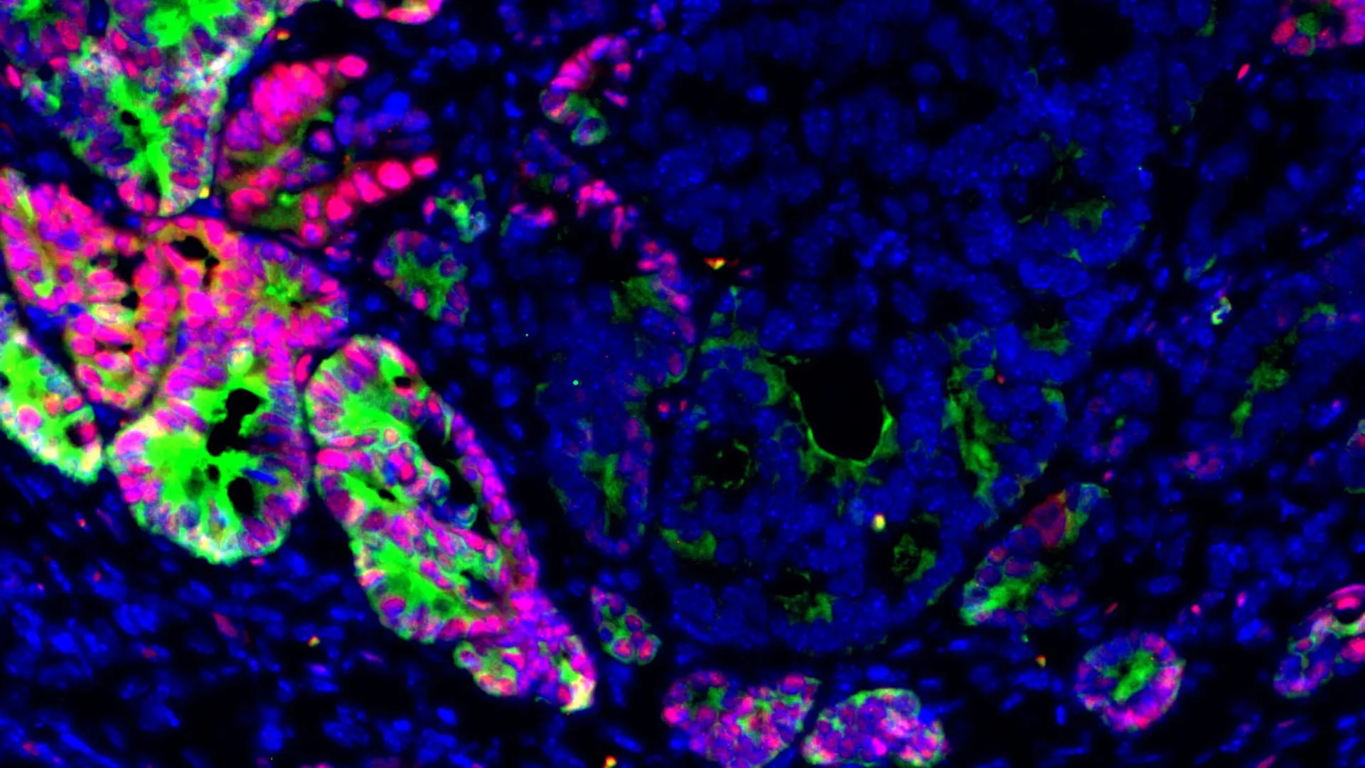
The biological mechanisms that allow colorectal cancer to migrate from the primary tumor site to the liver have long remained one of the most significant mysteries in oncology. However, a collaborative study led by researchers at Weill Cornell Medicine and the Massachusetts Institute of Technology (MIT) has identified a pivotal molecular factor—the transcription factor GATA6—that governs this transition. Published in the journal Cell Stem Cell on June 22, the findings reveal that the loss of GATA6 triggers a cascade of cellular changes, effectively stripping cancer cells of their specialized identity and reverting them to a highly adaptable, primitive state. This transformation, known as lineage plasticity, is what allows these cells to survive the arduous journey through the bloodstream and colonize the liver, the most common site for colorectal cancer metastasis.
Colorectal cancer (CRC) remains the third most common cancer diagnosed in both men and women in the United States and the second leading cause of cancer-related deaths worldwide. According to data from the American Cancer Society, approximately 153,000 new cases are diagnosed annually in the U.S. alone. While localized colorectal cancer has a high five-year survival rate of roughly 91%, that figure plummets to 15% once the disease has metastasized to distant organs. The liver is the primary destination for these migrating cells, largely due to the venous drainage system of the intestinal tract, which flows directly into the liver via the portal vein. Despite decades of genomic sequencing, researchers have struggled to find a consistent "metastasis gene" or mutation that explains why some tumors spread while others remain localized. This new research suggests the answer lies not in the DNA sequence itself, but in how the DNA is regulated.
The Search for the Metastatic Driver
For years, the scientific community focused on identifying specific genetic mutations—permanent changes to the DNA code—that might act as "on" switches for metastasis. However, comprehensive genomic studies frequently showed that primary colorectal tumors and their corresponding liver metastases shared almost identical mutation profiles. This led many to believe that the ability to spread was not a result of new mutations, but rather a result of epigenetic shifts—changes in gene expression that do not alter the underlying DNA sequence.
Dr. Norihiro Goto, an assistant professor of medicine in the Division of Gastroenterology & Hepatology at Weill Cornell and a co-lead author of the study, explained that the research team sought to identify these epigenetic "switches." They focused on GATA6, a transcription factor known to be essential for the development and maintenance of the intestinal lining. In a healthy gut, GATA6 acts as a molecular "identity keeper," ensuring that cells perform their specialized functions, such as nutrient absorption and mucus production. When GATA6 is present, cells are "locked" into their adult roles. When it is lost, that lock is broken.
The study involved an extensive analysis of patient samples and mouse models. The researchers observed that GATA6 levels were consistently lower in liver metastases compared to the original primary tumors. Furthermore, they found a direct correlation between low GATA6 expression and poor clinical outcomes in patients. This suggested that GATA6 loss was not just a byproduct of cancer progression, but a functional driver of the disease’s most lethal stage.
Innovative Organoid Modeling and the Chronology of Discovery
To capture the early, elusive stages of metastasis, the research team turned to cutting-edge organoid technology. Standard 2D cell cultures often fail to replicate the complex architecture and behavior of human tumors. In contrast, organoids are three-dimensional clusters of cells that mimic the structure and biological responses of real organs or tumors.
The researchers developed organoids derived from liver metastases and genetically engineered them to allow for the manipulation of GATA6. The chronological progression of the experiment involved several key phases:
- Initial Cultivation: Researchers grew colorectal cancer organoids that retained high levels of GATA6.
- Implantation: These organoids were implanted into the colons of mice to observe the development of primary tumors.
- Serial Selection: In a process known as "in vivo selection," the researchers harvested the most aggressive cells that managed to reach the liver and used them to create new organoids.
- Comparison: By repeating this cycle, the team could compare the genetic and epigenetic profiles of cells that stayed in the colon versus those that successfully colonized the liver.
This iterative process revealed that as the cancer cells became more adept at spreading, GATA6 expression progressively declined. When the researchers artificially deleted GATA6 from the primary tumor organoids, the results were striking: the frequency of liver metastasis in the mouse models increased significantly. Notably, the loss of GATA6 did not necessarily make the primary tumor grow faster or larger; instead, it specifically enhanced the cells’ ability to migrate and survive in the liver environment.
Lineage Plasticity: Reverting to a Fetal State
The most profound discovery of the study was the mechanism by which GATA6 loss promotes metastasis. The researchers found that without GATA6, colorectal cancer cells undergo a process called lineage plasticity. This is a state of cellular "fluidity" where the cells lose their specialized intestinal identity and adopt characteristics of fetal intestinal cells.
In a healthy body, this type of plasticity is a vital survival mechanism. For instance, during a major injury to the gut, cells may temporarily revert to a more primitive state to facilitate rapid tissue repair and wound healing. Once the repair is complete, they normally return to their specialized adult state. In the context of cancer, however, this process is hijacked. By entering a "fetal-like" state, cancer cells become highly adaptable, allowing them to survive the stresses of the circulatory system and the foreign environment of the liver.
A key marker of this transition is the loss of LGR5, a protein typically associated with adult intestinal stem cells. Previous research had hinted that LGR5-negative cells might be responsible for seeding metastases, but the reason for their appearance was unclear. The Weill Cornell and MIT study demonstrated that the loss of GATA6 is the specific trigger that shifts cells from an LGR5-positive state to an LGR5-negative, fetal-like state. These LGR5-negative cells are the "scouts" of the tumor, possessing the flexibility required to establish new colonies in distant organs.
Clinical Implications and Potential Biomarkers
The identification of GATA6 as a critical regulator of metastasis has immediate implications for the diagnosis and treatment of colorectal cancer. Currently, doctors rely heavily on the size of a tumor and its depth of invasion (the "T" in TNM staging) to determine a patient’s risk. However, many patients with small, early-stage tumors still go on to develop metastatic disease.
By measuring GATA6 levels in biopsy samples from primary tumors, clinicians may be able to more accurately predict which patients are at high risk for liver metastasis. A tumor with low GATA6 expression could serve as a "red flag," indicating that the cancer has already begun the process of cellular reshaping required for spread. This would allow for more aggressive monitoring and the earlier use of systemic therapies, such as chemotherapy or targeted biologics, even if the primary tumor appears small.
Dr. Saori Goto, an instructor in medicine at Weill Cornell and the study’s first author, emphasized that restoring GATA6 or its downstream pathways could represent a new therapeutic frontier. In laboratory experiments, the team found that restoring GATA6 activity effectively "re-specialized" the cancer cells, reducing their ability to form metastatic colonies.
Challenges and Future Directions
Despite the promise of these findings, translating them into a clinical treatment presents significant hurdles. Because the biological programs controlled by GATA6 are also used for normal tissue repair, any drug designed to block lineage plasticity must be highly selective. Interfering with these pathways globally could prevent the body from healing itself after injury or surgery.
The research team is now shifting its focus to identifying "vulnerabilities" that are unique to GATA6-deficient cells. If these cells have a specific weakness—perhaps a dependence on a certain nutrient or a vulnerability to a specific type of stress—it may be possible to target them without harming healthy tissue.
Furthermore, the team plans to investigate the role of the "tumor microenvironment." Metastasis is not just about the cancer cell; it is also about how the cell interacts with the immune system and the specific environment of the liver. Understanding how the liver "welcomes" these GATA6-deficient fetal-like cells could lead to therapies that make the liver less hospitable to cancer.
Dr. Omer H. Yilmaz, associate professor of biology at MIT and co-lead author, noted that this study marks a shift in how oncology views the progression of the disease. "We are moving beyond the idea that cancer is just a collection of mutations," Yilmaz stated. "We are beginning to see it as a failure of cellular identity."
The study was a massive undertaking supported by a wide array of institutions, including the National Institutes of Health (NIH), the Astellas Foundation, the Pew-Stewart Trust, and the Crohn’s & Colitis Foundation. As the research moves into preclinical phases, the ultimate goal remains clear: to transform colorectal cancer from a potentially fatal systemic disease into a manageable, localized condition by cutting off its ability to spread at the source. By stabilizing the molecular "identity" of cancer cells, scientists may finally be able to prevent the "identity crisis" that leads to metastasis.
