
 By Suherman Candra Maholtra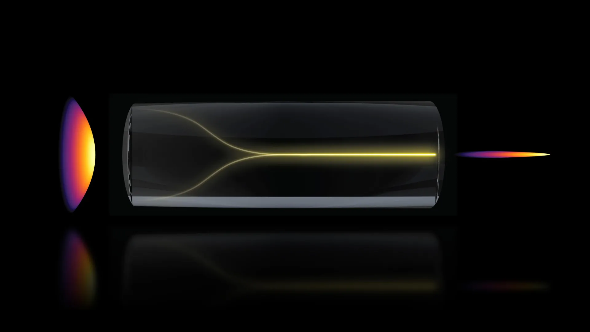
In a discovery that challenges long-standing assumptions in optical physics, a research team at the Massachusetts Institute of Technology (MIT) has identified a phenomenon where chaotic laser signals spontaneously reorganize into a highly focused "pencil beam." This breakthrough, detailed in a recent publication in Nature Methods, provides a transformative approach to biological imaging, enabling scientists to observe the human blood-brain barrier in three dimensions at speeds 25 times faster than current industry-standard techniques. By harnessing the self-organizing properties of light within multimode optical fibers, the researchers have developed a method that maintains high-resolution clarity while significantly increasing the depth of focus, offering a new window into how drugs interact with the brain’s most restrictive physiological defenses.
The Paradigm Shift in Nonlinear Optics
For decades, the consensus among optical physicists was that increasing the power of a laser traveling through a multimode optical fiber would inevitably lead to increased entropy. Multimode fibers are designed to carry high levels of energy, but because they allow light to travel along multiple paths simultaneously, the resulting output is typically a "speckle pattern"—a grainy, disordered distribution of light caused by internal interference and fiber imperfections. To correct this, engineers usually rely on complex "beam-shaping" components or digital algorithms to force the light into a usable state.
However, the MIT team, led by Assistant Professor Sixian You of the Department of Electrical Engineering and Computer Science (EECS), found that under specific conditions, the fiber does the work itself. As power is scaled toward the threshold of material damage, the light undergoes a nonlinear transition. Instead of shattering into further chaos, the electromagnetic waves interact with the molecular structure of the glass fiber, effectively "self-focusing" into a stable, narrow beam.
"The common belief in the field is that if you crank up the power in this type of laser, the light will inevitably become chaotic," said Professor You. "But we proved that this is not the case. We followed the evidence, embraced the uncertainty, and found a way to let the light organize itself into a novel solution for bioimaging."
Chronology of the Discovery: From Error to Innovation
The discovery began as an unexpected observation during routine stress tests of a "fiber shaper," a device the team had previously developed to control light propagation. Lead author Honghao Cao, an EECS graduate student, was tasked with gradually increasing the laser power to determine the physical limits of the multimode fiber.
In a typical experimental trajectory, researchers avoid high-power thresholds to prevent permanent damage to the equipment. However, as Cao pushed the intensity levels, the expected scattering did not occur. Instead, the team observed the light collapsing into a singular, intense point. This "pencil beam" emerged precisely when the system reached a critical power level where the nonlinearity of the glass began to counter the intrinsic disorder of the fiber.
To validate this finding, the team spent months identifying the exact parameters required to replicate the effect. They determined that two primary factors must be met: first, the laser must enter the fiber at a "zero-degree" angle of alignment, a requirement far stricter than those used in standard optical setups. Second, the power must reach a specific "nonlinearity threshold" where the light changes the refractive index of the fiber as it passes through. This balance creates a self-sustaining wave that resists the scattering effects of the fiber’s internal imperfections.
Solving the Depth-Resolution Tradeoff
In traditional microscopy, researchers face an inherent physical limitation known as the tradeoff between lateral resolution and depth of field. To see a cell in high detail (high resolution), the "waist" of the laser beam must be very thin. However, a thin waist usually means the beam diverges rapidly, resulting in a very shallow depth of focus. To image a thick 3D structure, like a blood vessel or a cluster of neurons, the microscope must take many individual 2D "slices" at different depths and then stack them together—a process that is both time-consuming and prone to motion artifacts.
The self-organized pencil beam bypasses this limitation. Because the beam is formed through nonlinear self-organization, it maintains its narrow, focused shape over a much longer distance than a standard Gaussian laser beam. This creates an extended "Rayleigh range," allowing the system to capture high-resolution data throughout a thick volume of tissue in a single pass.
Comparative testing revealed that this pencil beam is remarkably "clean." Many high-intensity laser systems produce "sidelobes"—secondary halos of light that blur the edges of an image. The MIT-developed beam, however, remains tightly concentrated, producing images with significantly fewer artifacts and higher contrast.
Applications in Neuropharmacology and the Blood-Brain Barrier
The most immediate and impactful application of this technology is the study of the human blood-brain barrier (BBB). The BBB is a highly selective semipermeable border of endothelial cells that prevents solutes in the circulating blood from non-selectively crossing into the central nervous system. While this protects the brain from toxins and pathogens, it also serves as a massive hurdle for drug delivery. Approximately 98% of small-molecule drugs and nearly 100% of large-molecule drugs fail to cross the BBB, making diseases like Alzheimer’s, ALS, and glioblastoma notoriously difficult to treat.
Using the new imaging technique, the MIT researchers, in collaboration with Professor Roger Kamm and colleagues at Harvard University and Beth Israel Deaconess Medical Center, were able to visualize 3D models of the human BBB with unprecedented speed. The system recorded images 25 times faster than current "gold-standard" multiphoton microscopy.
Crucially, the method is "label-free," meaning it does not require the use of toxic fluorescent dyes or genetic modifications to make cells visible. This allows scientists to watch individual cells absorb proteins and drugs in real-time under natural conditions.
"The pharmaceutical industry is especially interested in using human-based models to screen for drugs that effectively cross the barrier, as animal models often fail to predict what happens in humans," explained Roger Kamm, the Cecil and Ida Green Distinguished Professor of Biological and Mechanical Engineering at MIT. "That this new method doesn’t require the cells to have a fluorescent tag is a game-changer. For the first time, we can now visualize the time-dependent entry of drugs into the brain."
Supporting Data and Technical Performance
The technical specifications of the self-organized beam highlight its superiority over existing methods:
- Imaging Speed: The pencil beam approach achieves a 25-fold increase in volumetric imaging speed compared to traditional point-scanning systems.
- Depth of Focus: The self-organized beam maintains a cellular-level resolution (approx. 1 micrometer) over a depth of focus that is several times longer than what is physically possible with standard lenses of the same aperture.
- Stability: Despite the high power levels required to trigger the effect, the beam remains stable over long durations, allowing for the "time-resolved" tracking of molecular targets.
- Accessibility: Because the effect is intrinsic to the physics of the fiber and the laser, it can be implemented using standard optical components, potentially lowering the cost of high-end bioimaging.
Sarah Spitz, a postdoc and co-author on the study, noted that the implications extend far beyond the brain. "This approach enables time-resolved tracking of diverse compounds and molecular targets across engineered tissue models, providing a powerful tool for biological engineering," she stated.
Future Outlook and Institutional Impact
The discovery of self-organizing light in multimode fibers opens a new chapter in the study of nonlinear dynamics. The MIT team plans to delve deeper into the underlying physics to determine if this effect can be further optimized or applied to different types of optical media.
From a clinical perspective, the ability to rapidly screen drug candidates in human-derived tissue models could significantly shorten the timeline for neurodegenerative disease research. By identifying exactly which cell types internalize a drug and at what rate, researchers can refine dosages and delivery mechanisms before moving to expensive and risky human trials.
The research was a multi-disciplinary effort involving the MIT Research Laboratory for Electronics (RLE), the Department of Biological Engineering, and clinical partners at Harvard. Funding for the project was provided by a diverse coalition of organizations, including the National Science Foundation (NSF), the Silicon Valley Community Foundation, the Diacomp Foundation, and the Claude E. Shannon Award.
As the team moves toward commercializing or broadly distributing this technology, the focus will remain on transitioning the "pencil beam" from a laboratory curiosity into a standard tool for the global scientific community. By turning what was once considered a "hassle" of optical engineering into a self-solving solution, the MIT researchers have paved the way for a faster, clearer, and more efficient era of biological discovery.
