
 By George Ongkojoyo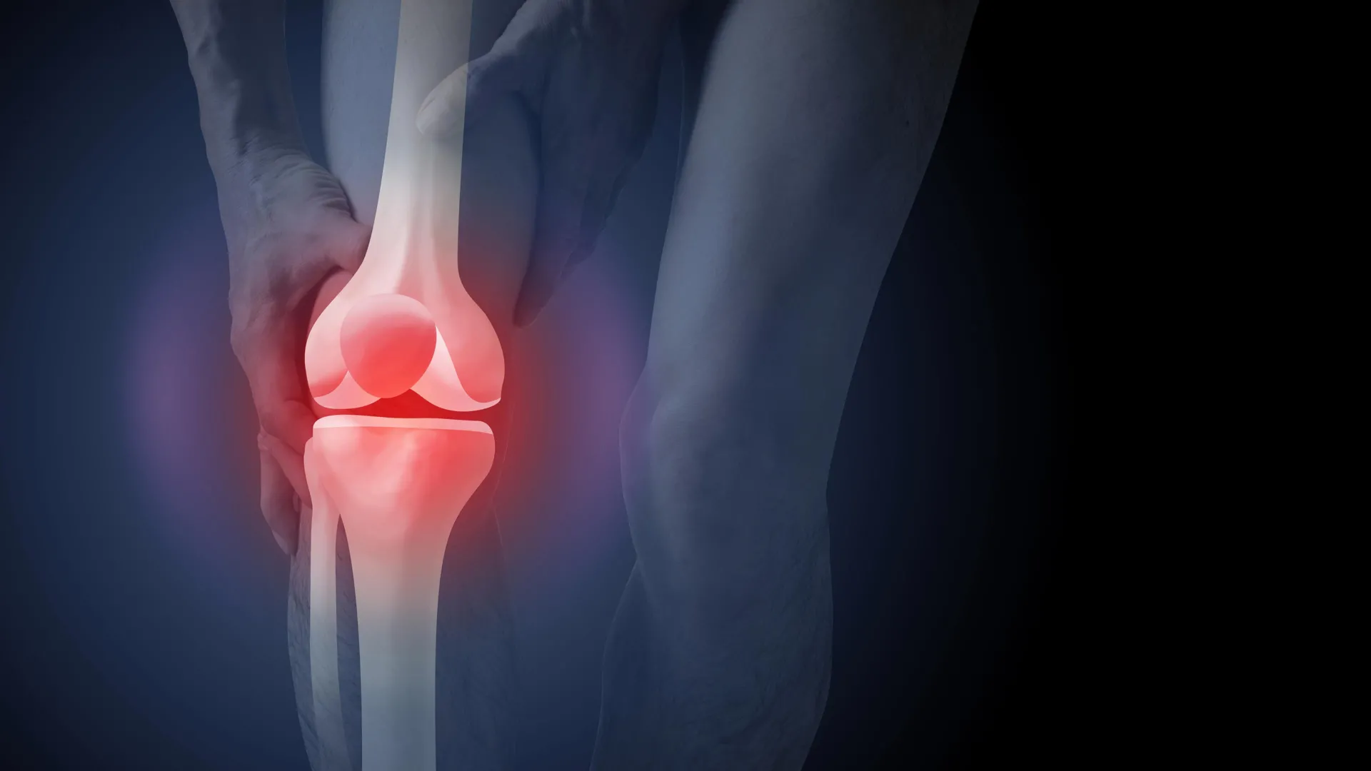
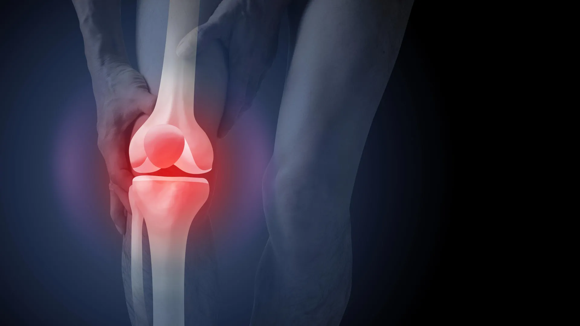
A groundbreaking study published in the journal PLOS Medicine has revealed that the routine use of X-rays to diagnose knee osteoarthritis may be doing more harm than good for patient outcomes and healthcare sustainability. Despite long-standing clinical guidelines advising against imaging for initial diagnosis, nearly half of all new patients in Australia presenting with knee pain are referred for X-rays. The research suggests that viewing these images fundamentally alters a patient’s perception of their condition, leading to increased fear of movement, heightened anxiety about joint "damage," and a significantly higher desire for invasive surgical interventions that may not be medically necessary.
The study, which involved 617 participants across Australia, highlights a critical disconnect between evidence-based medical standards and current clinical practice. While osteoarthritis is a leading cause of disability worldwide, the reliance on structural imaging rather than clinical symptoms is contributing to a ballooning healthcare bill, with osteoarthritis-related imaging costing the Australian health system approximately A$104.7 million annually. Furthermore, the broader economic impact of the condition, driven largely by joint replacement surgeries, reached a staggering $3.7 billion in the 2020–21 fiscal year.
The Psychological Impact of Imaging: A Randomized Controlled Trial
The core of the new research focused on how the method of diagnosis influences a patient’s mindset and subsequent health choices. Researchers employed a randomized controlled trial design, using video vignettes to simulate a consultation with a General Practitioner (GP). Participants were divided into three distinct groups: one group received a clinical diagnosis based solely on age and symptoms; the second group received an X-ray-based diagnosis but did not see the images; and the third group received an X-ray-based diagnosis and was shown the radiographic evidence of their joint changes.
The results were stark. Participants who were shown their X-ray images reported a 36% higher perceived need for total knee replacement surgery compared to those who received a clinical diagnosis without imaging. This group also exhibited a marked increase in "fear-avoidance" beliefs, assuming that physical activity and exercise would cause further "wear and tear" on the joint. Paradoxically, the study found that patients were slightly more satisfied with a diagnosis that included an X-ray, suggesting a deeply ingrained cultural belief that "seeing is believing" when it comes to internal pain.
Medical experts suggest this satisfaction is a "double-edged sword." While patients feel validated by seeing a physical abnormality on a screen, that validation often morphs into a conviction that their joint is "broken" or "worn out," leading them to reject effective non-surgical treatments like physiotherapy and weight management in favor of the perceived "fix" of surgery.
Deconstructing the "Wear and Tear" Myth
For decades, the prevailing narrative around osteoarthritis has been that it is a mechanical failure—a simple case of the cartilage wearing away like the tread on a tire. However, modern medical science paints a much more complex biological picture. Osteoarthritis is now understood as a condition involving the entire joint environment, including the bone, ligaments, muscles, and the synovial fluid. It is a process where the joint is working overtime to repair itself in response to various stressors, including age, weight, and previous injury.
Crucially, research has consistently shown that the severity of structural changes visible on an X-ray—such as bone spurs or narrowed joint spaces—does not correlate well with the level of pain or disability a person experiences. Many individuals with "severe" X-ray results live active, pain-free lives, while others with "normal" X-rays experience debilitating symptoms. Because imaging cannot predict symptom progression or pain levels, the Australian Commission on Safety and Quality in Health Care, along with international bodies like the National Institute for Health and Care Excellence (NICE) in the UK, explicitly recommend against routine X-rays for patients over the age of 45 who present with typical symptoms.
The Financial Burden on the Australian Healthcare System
The over-reliance on imaging is not merely a clinical issue but a significant economic one. According to data from the Australian Institute of Health and Welfare (AIHW), the financial footprint of osteoarthritis is expanding rapidly as the population ages.
- Imaging Costs: At A$104.7 million per year, the cost of knee and hip imaging represents a significant portion of primary care expenditure. Much of this is considered "low-value care," providing little to no benefit to the patient’s eventual recovery path.
- Surgical Expenditure: In 2021–22, more than 53,000 Australians underwent knee replacement surgery. While these procedures are life-changing for those with end-stage disease who have exhausted all other options, the $3.7 billion annual cost of hospital services for osteoarthritis suggests a system heavily weighted toward high-cost surgical solutions.
- Productivity Loss: Beyond direct medical costs, the condition contributes to billions in lost productivity and early retirement, as patients who fear movement due to their X-ray results are less likely to remain in the workforce.
A Chronology of Diagnostic Standards
The shift away from imaging has been a gradual process in the medical community, though public perception has lagged behind.
- 1950s–1990s: The Kellgren-Lawrence grading system established X-rays as the "gold standard" for identifying osteoarthritis, focusing almost entirely on structural changes.
- 2000s: Longitudinal studies began to reveal the "pain-radiograph gap," showing that many people with radiographic osteoarthritis had no symptoms.
- 2010s: Major health organizations began issuing "Choosing Wisely" recommendations, urging doctors to avoid unnecessary imaging for musculoskeletal pain.
- 2023–2024: New clinical care standards in Australia emphasize "clinical diagnosis," defined as a patient aged 45 or older who experiences activity-related joint pain and has either no morning stiffness or stiffness lasting less than 30 minutes.
The Risks of Unnecessary Surgery
The study’s finding that X-rays drive a 36% increase in surgical demand is particularly concerning given the risks associated with total knee arthroplasty (TKA). While generally successful, TKA is major surgery involving significant rehabilitation. Potential adverse events include deep vein thrombosis (blood clots), pulmonary embolism, surgical site infections, and chronic post-surgical pain.
Statistically, approximately 10% to 20% of patients who undergo knee replacement remain dissatisfied with the results, often because the surgery did not address the underlying functional issues or because their expectations were not aligned with the reality of recovery. By bypassing conservative management—such as targeted exercise programs like GLAD (Good Life with osteoArthritis in Denmark)—patients may be taking on surgical risks without first attempting lower-risk, high-reward interventions.
Expert Reactions and Policy Implications
While the study authors emphasize that changing clinical practice is challenging, the implications for policy are clear. There is an urgent need for "de-implementation" strategies to reduce the number of unnecessary X-ray referrals.
Inferred reactions from the broader medical community suggest a two-pronged approach. First, GPs require better support and longer consultation times to explain a clinical diagnosis to patients, ensuring the patient feels heard without needing a "picture" of their bone. Second, public health campaigns are needed to rebrand osteoarthritis from a "wear and tear" disease to a "long-term joint health" issue that responds well to movement.
Advocates for value-based healthcare argue that redirecting even a fraction of the $104.7 million spent on imaging toward subsidized physiotherapy or weight management programs could yield far better long-term outcomes for the Australian population.
Future Outlook: Moving Toward Symptom-Based Care
The findings of this study serve as a wake-up call for both clinicians and patients. For the individual, the message is one of empowerment: a diagnosis of osteoarthritis is not a death sentence for an active lifestyle, nor does a "scary" X-ray image accurately predict one’s future mobility.
The medical community is increasingly moving toward a holistic model of care that prioritizes education, exercise, and weight control as the first line of defense. By reducing the emphasis on "damage" seen on X-rays, healthcare providers can help patients focus on what they can do—strengthening the muscles around the joint and improving overall function—rather than what they fear is "broken" inside.
As the Australian government continues to look for ways to curb rising healthcare costs, addressing the "low-value care" of routine osteoarthritis imaging remains a low-hanging fruit. Reducing unnecessary X-rays not only saves money and reduces radiation exposure but, as this study proves, it may also be the key to preventing thousands of patients from undergoing surgery they might never have needed.
