
 By Suherman Candra Maholtra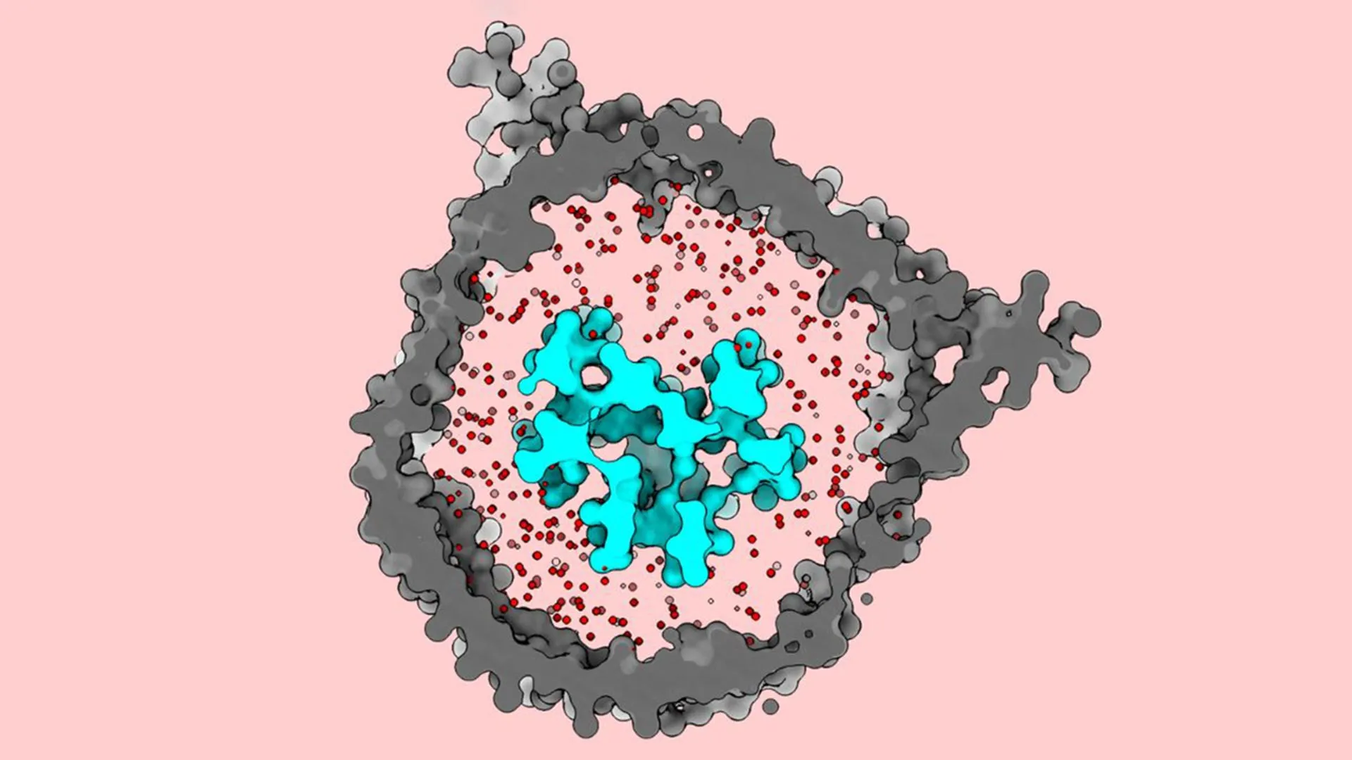
The landscape of modern oncology has been fundamentally reshaped over the last decade by the advent of T cell immunotherapy, a revolutionary approach that harnesses the body’s own immune system to identify and eradicate malignant cells. While these therapies have achieved remarkable success in treating certain blood cancers, such as leukemia and lymphoma, they have frequently encountered a "glass ceiling" when applied to solid tumors and various other cancer types. For years, the scientific community has operated these therapies with an incomplete blueprint, utilizing the T cell receptor (TCR) as a primary tool without fully grasping the molecular mechanics of its activation. However, a landmark study conducted by researchers at The Rockefeller University, recently published in Nature Communications, has finally illuminated the structural secrets of the TCR, revealing a "jack-in-the-box" mechanism that could hold the key to the next generation of cancer treatments.
The Structural Mystery of the T Cell Receptor
At the heart of the adaptive immune response is the T cell, a specialized white blood cell tasked with patrolling the body for signs of infection or malignancy. The T cell’s "eyes" are its receptors—complex protein structures embedded within the cell membrane. These receptors are responsible for scanning the surfaces of other cells, looking for foreign peptides (antigens) presented by human leukocyte antigen (HLA) complexes. When a TCR recognizes a specific antigen, it triggers a signaling cascade that transforms the T cell from a dormant surveyor into an aggressive killer.
Despite the clinical importance of this process, the earliest physical changes that occur when a TCR encounters an antigen have remained one of the great enigmas of molecular biology. Understanding these changes is not merely an academic exercise; it is a clinical necessity. Because scientists did not fully understand how the TCR "switches on," they lacked the precision tools required to fine-tune the receptor’s sensitivity. This has led to a major hurdle in immunotherapy: many T cell treatments are either not sensitive enough to detect low levels of cancer antigens or too aggressive, leading to the destruction of healthy tissue.
A Paradigm Shift in Molecular Imaging
The breakthrough at Rockefeller University was led by Thomas Walz, a world-renowned expert in cryo-electron microscopy (cryo-EM), and Ryan Notti, an instructor in clinical investigation and a physician at Memorial Sloan Kettering Cancer Center. Their collaboration sought to resolve a glaring contradiction in existing scientific literature. Previous structural studies, which utilized cryo-EM to visualize the TCR, had depicted the receptor as an "open" and extended structure even when it was inactive. This data suggested that the receptor did not undergo significant shape changes upon binding to an antigen—a finding that baffled biologists who knew that a mechanical signal must be sent from the outside of the cell to the inside to initiate an immune response.
Notti and Walz hypothesized that the previous studies were flawed not by their technology, but by their methodology. Most earlier attempts to image the TCR relied on detergents to extract the protein complex from the cell membrane. While detergents make proteins easier to handle in a laboratory setting, they strip away the lipid bilayer—the fatty "skin" of the cell. The Rockefeller team suspected that without this natural membrane to hold it in place, the TCR was essentially falling apart or springing open prematurely, leading to a false representation of its dormant state.
Reconstructing the Native Milieu: The Nanodisc Approach
To observe the TCR in its true form, the researchers employed an innovative technique involving "nanodiscs." These are microscopic, disc-shaped sections of a synthetic membrane that mimic the natural environment of a living cell. By carefully assembling the TCR’s eight constituent proteins within these nanodiscs, the team was able to recreate the receptor’s native environment with unprecedented accuracy.
This process was fraught with biochemical challenges. The TCR is a notoriously unstable complex, and ensuring that all eight proteins were correctly oriented within the lipid bilayer required meticulous precision. Furthermore, the team did not use a generic model lipid; they developed a specific lipid mixture that mirrored the unique chemical composition and thickness of a real T cell membrane. "We can change the biochemical composition, the thickness of the membrane, the tension and curvature—all kinds of parameters that we know have an influence on the embedded protein," Walz explained, highlighting the level of customization required for the study.
The Jack-in-the-Box Revelation
When the researchers finally visualized the TCR within the nanodiscs using cryo-EM, the results were transformative. Contrary to previous studies, the dormant TCR was found to be tightly packed and compact. It did not sit in an open position; rather, it remained folded down, restrained by the surrounding lipid membrane.
The most significant discovery occurred when the receptor encountered an antigen-presenting molecule. Upon binding, the TCR underwent a dramatic conformational change, springing upward and outward. This "jack-in-the-box" motion provides the mechanical force necessary to transmit a signal through the cell membrane and into the interior of the T cell. The lipid membrane acts as the "lid" of the box, keeping the receptor compressed until the correct antigen provides the "key" to release it.
This finding explains why previous detergent-based studies were misleading. By removing the membrane, earlier researchers had effectively "opened the lid," allowing the receptor to spring into its active shape without an antigen present. The Rockefeller study proves that the membrane is not just a passive container for the TCR, but an active participant in its regulation.
Clinical Implications for Sarcomas and Solid Tumors
The motivation for this research was deeply personal for Ryan Notti. As an oncologist at Memorial Sloan Kettering, Notti treats patients with sarcomas—rare and often aggressive cancers that develop in the bones and soft tissues. While immunotherapy has been a miracle for some, it has largely failed sarcoma patients.
"The T cell receptor is really the basis of virtually all oncological immunotherapies, so it’s remarkable that we use the system but really have had no idea how it actually works," Notti noted. For patients with solid tumors, the immune system often fails to recognize cancer cells because the antigens they present are too similar to healthy cells, or they are presented in such low quantities that the TCR fails to "spring."
With this new understanding of the TCR’s mechanical activation, researchers can now begin to "tune" the receptor. By re-engineering the TCR to have a lower activation threshold, scientists could theoretically make T cells more sensitive to the faint signals emitted by sarcomas. Conversely, for autoimmune diseases where T cells mistakenly attack the body, treatments could be designed to stabilize the "closed" state of the receptor, preventing unwanted activation.
Data Analysis and Future Research Trajectories
The data produced by Walz’s lab provides a high-resolution roadmap that was previously non-existent. By analyzing the specific points of contact between the TCR and the HLA-antigen complex, researchers can now use computational modeling to predict how different mutations in the receptor will affect its "spring" mechanism.
Supporting data from the study suggests that the thickness and lipid composition of the membrane significantly influence the energy required for the TCR to transition from a closed to an open state. This opens a new frontier in "lipid-targeted" therapies, where the environment of the T cell itself might be modified to enhance its cancer-fighting capabilities.
Furthermore, the implications extend to vaccine development. Effective vaccines work by training the TCR to recognize specific viral or bacterial antigens. The Rockefeller structures allow vaccine designers to see the refined details of these interactions, potentially leading to vaccines that elicit a more robust and precise T cell response. This could be particularly relevant for developing vaccines against rapidly mutating viruses or pathogens that have historically evaded the immune system.
A New Era of Precision Immunotherapy
The study’s publication in Nature Communications marks a pivotal moment in the transition from "black box" medicine to precision engineering in oncology. Thomas Walz, reflecting on the decades of work in his laboratory, described this as "some of the most important work to ever come out of my lab."
The ability to visualize the TCR in its native state solves a fundamental biological mystery and provides a tangible path forward for clinical applications. As researchers move toward "Next-Generation Immunotherapy," the focus will shift from simply activating the immune system to precisely controlling it. The "jack-in-the-box" mechanism provides the blueprint for this control, offering hope that the successes seen in blood cancer treatments can eventually be replicated across the entire spectrum of oncological disease.
In the coming years, the research community expects to see a surge in "adoptive T cell therapies" that utilize these structural insights. By customizing the TCR’s sensitivity, clinicians may finally be able to break through the barriers that have protected solid tumors for so long, turning the tide in the fight against some of the most challenging forms of cancer. This study stands as a testament to the power of basic science—proving that by understanding the smallest molecular gears of life, we can find the leverage needed to move the entire field of medicine forward.
