
 By Gabriel Paijo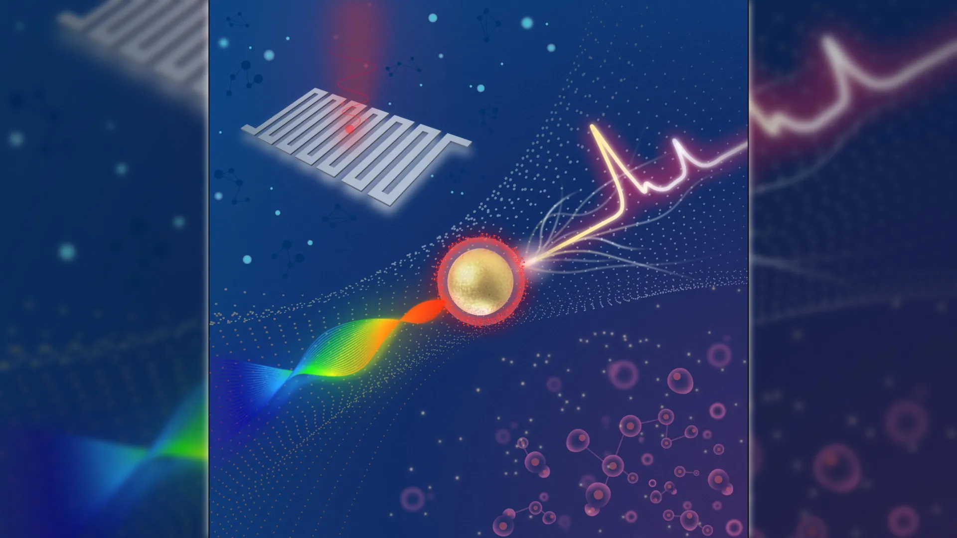
A multidisciplinary research team led by the Michigan State University Institute for Quantitative Health Science and Engineering (IQ) has successfully engineered a compact Raman imaging system capable of distinguishing between cancerous and healthy tissue with unprecedented sensitivity. This technological leap, detailed in the latest issue of the journal Optica, utilizes a combination of advanced quantum sensing and nanoparticle engineering to overcome the traditional limitations of molecular imaging. By detecting tumor-specific biomarkers at concentrations previously thought unreachable by portable devices, the system paves the way for real-time, intraoperative cancer screening and significantly faster diagnostic workflows.
The Evolution of Cancer Diagnostics: From Pathology to Real-Time Molecular Imaging
For over a century, the gold standard for cancer diagnosis has remained largely unchanged: the histological examination of tissue biopsies. This process involves the physical removal of tissue, followed by chemical staining—most commonly with hematoxylin and eosin (H&E)—and a meticulous review by a trained pathologist. While highly accurate, this method is inherently retrospective and time-consuming. Patients often wait days or weeks for results, a delay that can increase psychological distress and, in aggressive cases, allow the disease to progress.
The emergence of Raman spectroscopy offered a potential alternative. Named after Sir C.V. Raman, who discovered the phenomenon in 1928, the technique involves shining a laser on a sample and measuring the "inelastic scattering" of photons. This scattering provides a unique "chemical fingerprint" of the molecules within the sample. However, Raman signals are notoriously weak, often obscured by background fluorescence and electronic noise. To solve this, scientists developed Surface-Enhanced Raman Scattering (SERS), which uses metallic nanoparticles to amplify these signals by several orders of magnitude.
Despite these advancements, bringing Raman imaging into the operating room or a local clinic has been hindered by the size and cost of the equipment. Traditional systems require bulky spectrometers and high-powered cameras that are sensitive to vibration and temperature changes. The breakthrough by Zhen Qiu and his team at Michigan State University addresses these hurdles by reimagining the architecture of the Raman system itself.
Technical Architecture: Synergizing Swept-Source Lasers and Superconducting Nanowire Detectors
The core of the MSU system’s superiority lies in its unique hardware configuration. Rather than using a traditional dispersive spectrometer, which spreads light across a fixed sensor, the team employed a "swept-source" Raman architecture. In this setup, a laser rapidly changes its wavelength across a specific range during the analysis. This approach allows the system to collect light more efficiently and simplifies the optical path, making the device significantly more compact.
The most critical component of the system, however, is the Superconducting Nanowire Single-Photon Detector (SNSPD). Developed in collaboration with the industry firm Quantum Opus, the SNSPD is a quantum-grade sensor that operates at near-zero electrical resistance. When a single photon hits the superconducting wire, it creates a "hotspot" that temporarily disrupts the superconductivity, generating a measurable voltage pulse.
This mechanism allows the MSU system to detect individual particles of light with nearly zero background noise. According to the published data, the system can identify Raman signals that are approximately four times weaker than those detectable by the highest-grade commercial Raman systems currently on the market. This sensitivity is measured at the femtomolar level—a scale so small it represents one-quadrillionth of a mole—allowing for the detection of tumor markers even when they are present in extremely low concentrations during the earliest stages of cancer development.
Experimental Validation: Targeting the CD44 Protein
To demonstrate the system’s clinical utility, the researchers focused on detecting CD44, a cell-surface glycoprotein that is overexpressed in a wide variety of cancers, including breast, colon, and prostate tumors. CD44 is frequently used as a marker for cancer stem cells, which are believed to be responsible for tumor initiation, metastasis, and resistance to chemotherapy.
The team engineered SERS nanoparticles coated with hyaluronan acid, a natural ligand that specifically binds to the CD44 receptor. The experimental process followed a specific chronology:
- Nanoparticle Synthesis: Gold nanoparticles were functionalized with Raman-active molecules and then encapsulated in a hyaluronan shell.
- In Vitro Testing: The system was first tested on cultured breast cancer cell lines. The imaging platform successfully highlighted the cells by detecting the unique Raman "fingerprint" of the nanoparticles attached to their surfaces.
- Ex Vivo Tissue Analysis: The researchers then applied the nanoparticles to mouse tumor samples and compared them to healthy control tissues.
- Data Processing: The system automatically processed the incoming signals, generating a heat map that clearly delineated the boundaries between malignant and healthy tissue.
The results were stark. The SERS signals were heavily concentrated in the tumor regions, while the healthy tissues showed almost no background interference. This high "signal-to-background" ratio is essential for clinical use, as it reduces the likelihood of false positives—a common problem with current imaging technologies like PET or MRI, which can sometimes flag inflammation as cancer.
Performance Metrics and Comparative Analysis
The MSU team’s findings highlight a significant shift in the performance envelope of Raman systems. In comparative benchmarks, the new system outperformed traditional charge-coupled device (CCD) based systems in several key areas:
- Sensitivity Threshold: The SNSPD-equipped system reached a detection limit in the femtomolar range, whereas standard commercial systems typically struggle below the picomolar range.
- Dynamic Range: The system demonstrated the ability to maintain accuracy across a wide range of signal intensities, which is crucial when imaging heterogeneous tumors that may have varying levels of biomarker expression.
- Form Factor: By using a fiber-coupled configuration, the researchers eliminated the need for free-space optics, which are prone to misalignment. This allows the system to be miniaturized into a portable unit roughly the size of a desktop computer, with the potential for further reduction into a handheld wand.
"Traditional methods are time-consuming and labor-intensive," noted Zhen Qiu. "While our system would not immediately replace the gold standard of pathology, it serves as a rapid screening tool that provides immediate feedback."
Broader Implications for Surgical Oncology and Personalized Medicine
The potential applications for this technology extend far beyond the initial laboratory results. One of the most promising uses is in "margin assessment" during cancer surgery. When a surgeon removes a tumor, they aim to take a small amount of healthy tissue around it—the "margin"—to ensure no cancer cells are left behind. Currently, surgeons must often wait for a "frozen section" analysis from a pathologist while the patient remains under anesthesia. If the margins are found to be "positive" (containing cancer), additional tissue must be removed, or the patient may require a second surgery later.
A compact, high-sensitivity Raman system could allow a surgeon to "scan" the surgical cavity in real-time. If the system detects the Raman signature of the SERS nanoparticles, the surgeon would know immediately that residual cancer cells are present, allowing for more precise and complete tumor removal in a single session.
Furthermore, the system’s adaptability makes it a candidate for personalized medicine. By changing the targeting molecule on the nanoparticle—swapping hyaluronan for antibodies that target HER2 (breast cancer) or PSA (prostate cancer)—the same hardware can be used to detect a wide array of different malignancies. The team is also exploring "multiplexing," a technique where different nanoparticles, each with a unique Raman signature, are used simultaneously to track multiple biomarkers at once. This would provide a comprehensive molecular profile of a patient’s tumor in minutes.
Path to Clinical Integration and Future Research
Despite the success of the current prototype, the transition from a laboratory setting to a clinical environment requires several additional steps. The research team has outlined a roadmap for the next three to five years:
- Increasing Readout Speed: Current imaging speeds are sufficient for small samples but need to be accelerated for large-scale tissue scanning. The team is investigating faster laser sources, such as Vertical-Cavity Surface-Emitting Lasers (VCSELs), to reduce scan times.
- Refining the Cooling System: SNSPDs require cryogenic temperatures to maintain their superconducting state. While the current system uses a compact cryostat, the team is working with partners like Quantum Opus to further streamline the cooling hardware for better portability.
- Human Clinical Trials: Following the successful mouse model studies, the team plans to move toward human tissue samples and, eventually, pilot clinical trials to validate the system’s safety and efficacy in a hospital setting.
The project has already garnered attention from the medical community and industry stakeholders. The collaboration with Quantum Opus highlights a growing trend of "quantum biology," where tools originally developed for quantum computing and deep-space communication are repurposed to solve complex biological problems.
Conclusion: A New Frontier in Medical Imaging
The development of the compact Raman imaging system at Michigan State University represents a convergence of nanotechnology, quantum physics, and oncology. By achieving femtomolar sensitivity in a compact format, Zhen Qiu and his colleagues have addressed the primary barriers that have kept Raman spectroscopy on the fringes of clinical practice for decades.
As the technology matures, it promises to reduce the "diagnostic gap"—the time between the first suspicion of cancer and the commencement of treatment. By providing clinicians with a high-resolution, molecular-level view of tissue in real-time, this system could significantly improve survival rates through earlier detection and more accurate surgical interventions. In the landscape of modern medicine, where every hour counts in the fight against cancer, such a leap in diagnostic speed and sensitivity is not merely an incremental improvement; it is a transformative shift toward a future of faster, more precise, and more accessible healthcare.
