
 By Budi Oetomo Narendra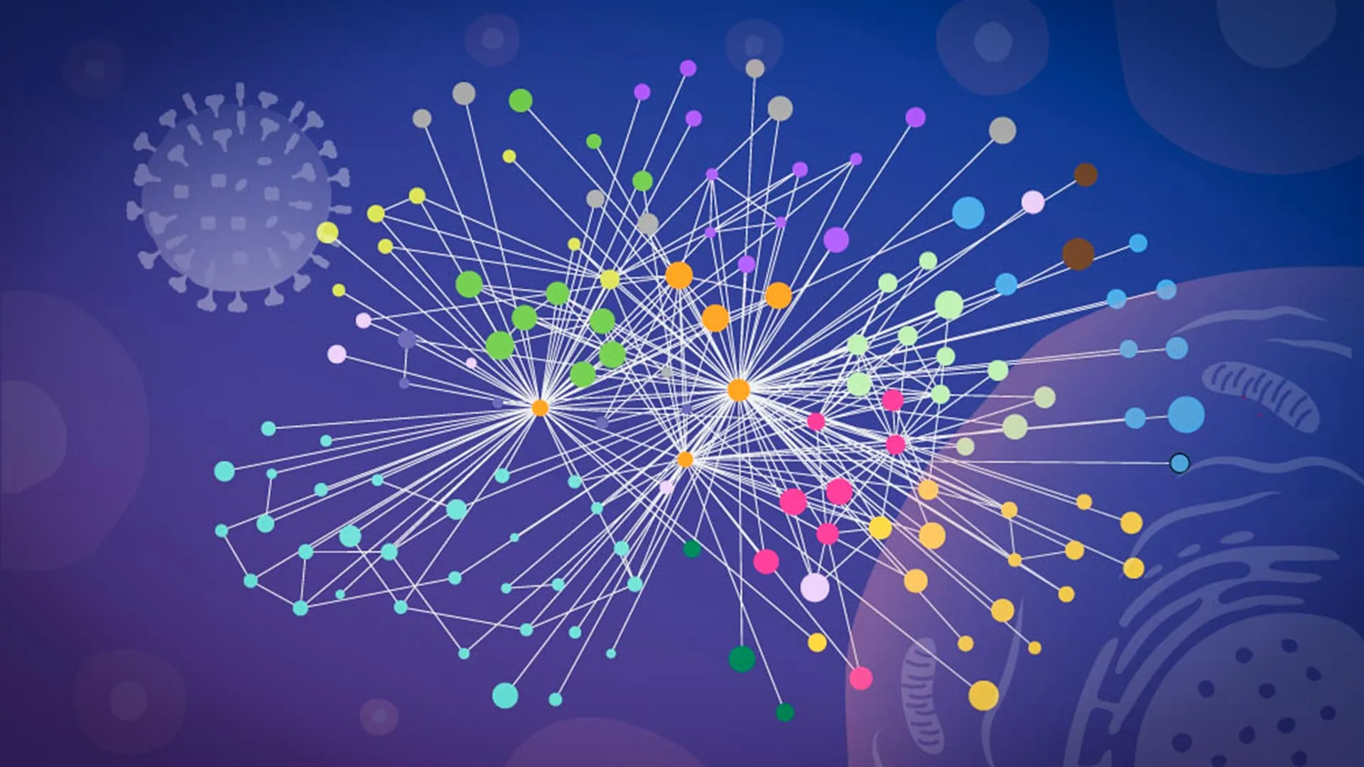
The Persistent Global Threat of Influenza A
Influenza, commonly known as the flu, remains one of the most significant public health challenges worldwide. Seasonal influenza outbreaks cause between 3 to 5 million cases of severe illness and are linked to as many as 290,000 to 650,000 respiratory deaths each year, according to the World Health Organization (WHO). Among the various types, influenza A viruses are particularly concerning due to their capacity for rapid evolution and their role in driving multiple pandemics throughout history.
The most devastating of these was the 1918 Spanish Flu pandemic, caused by an H1N1 influenza A virus, which is estimated to have killed between 50 and 100 million people globally – far exceeding the death toll of World War I. Subsequent pandemics include the 1957 Asian Flu (H2N2), the 1968 Hong Kong Flu (H3N2), and the 2009 Swine Flu (H1N1pdm09), each causing widespread illness and mortality. The recurring nature of influenza pandemics underscores the urgent need for a deeper understanding of how these viruses operate at a molecular level, enabling scientists to anticipate, prevent, and treat future outbreaks more effectively.
Influenza A viruses are RNA viruses with a segmented genome, meaning their genetic material is divided into several pieces. This characteristic allows for genetic reassortment – a process where different virus strains can swap genetic segments if they co-infect the same cell. This genetic mixing, along with continuous antigenic drift (small changes in viral surface proteins), allows influenza A to constantly evade the human immune system, necessitating annual vaccine reformulations and posing a continuous challenge for public health authorities.
Unlocking the Viral Blueprint: Unprecedented Insights into Cellular Hijacking
Upon entering a host cell, the influenza A virus initiates a sophisticated takeover. It releases its RNA genome, which contains the instructions for synthesizing a small but potent set of viral proteins. These proteins then spread throughout the host cell, systematically redirecting its complex molecular machinery to serve the virus’s replication needs, transforming the cell into a factory for new viral particles.
A clearer, more detailed view of this cellular subversion is critical for the development of more effective antiviral drugs and improved flu vaccines. To achieve this, researchers require precise knowledge of which viral proteins interact directly with human proteins, the exact locations of these crucial contacts within the cell, and the specific mechanisms by which the virus exploits these interactions to support its own propagation.
The new study, led by EMBL Hamburg’s Jan Kosinski and conducted in collaboration with Boris Bogdanow and Fan Liu at FMP Berlin, marks a significant leap forward. It represents the first large-scale mapping of direct contacts between influenza and human proteins inside intact infected cells, providing an unparalleled resolution. Furthermore, the structural detail gleaned from this research was precise enough to allow the scientists to computationally model how the interacting proteins physically fit together, offering a three-dimensional perspective on these critical molecular interfaces.
"Our work provides a new way to study flu-host interactions in their native context and with structural insight," explained Jan Kosinski, Group Leader at EMBL Hamburg and the Centre for Structural Systems Biology (CSSB). "The current results are a snapshot of a moment during infection, and it opens the door to studying flu-host interactions across the entire infection cycle." This initial "snapshot" lays foundational groundwork for future investigations that could potentially map the dynamic interplay throughout the full course of infection.
Overcoming Methodological Barriers: Preserving the Cellular Battlefield
Tracking the intricate web of protein-protein interactions during an active viral infection has historically been an extremely difficult scientific endeavor. Many earlier studies relied heavily on traditional biochemical techniques that mandated the destruction of the cell’s integrity – scientists had to break cells open, or "lyse" them, before they could measure protein contacts.
This destructive process, however, introduces significant limitations and potential artifacts. Once the delicate internal compartments of a cell are disrupted, proteins that were naturally separated by membranes or spatial arrangements within the living cell can come into artificial contact in the laboratory environment. Conversely, weak, transient, or highly localized interactions that are crucial to viral processes might be lost or disappear entirely once the cellular environment is compromised. Consequently, researchers often struggled to definitively determine which protein connections truly existed and were functionally relevant during the actual infection within a living cell.
The breakthrough came with the development of a specialized version of cross-linking mass spectrometry (XL-MS), a well-established technique for mapping protein contacts, specifically tailored for virus-infected cells. "This is when we learned that our collaborators, Boris Bogdanow and Fan Liu, at FMP Berlin had developed a specialized version of cross-linking mass spectrometry (XL-MS), a long-established technique for mapping protein contacts, tailored specifically to virus-infected cells," Kosinski noted.
Cross-linking mass spectrometry works by chemically "freezing" protein interactions in situ within the intact cell. Small chemical linkers are introduced that form covalent bonds between proteins that are in close proximity. The cells are then lysed, and the cross-linked protein complexes are analyzed using mass spectrometry. This specialized method provided the crucial advantage the team needed: it allowed them to capture interactions that occur only briefly or within particular regions of an infected cell, providing a far more accurate representation of the molecular events unfolding during infection.
Boris Bogdanow, now a Junior Research Group Leader at the Institute of Virology, Charité – Universitätsmedizin Berlin, elaborated on the technique’s power: "XL-MS allows us to capture protein-protein interactions directly in infected intact cells, while also providing structural information about how these interactions are happening. This gives us insight into the interface between the virus and the human cell and may, through structural modeling, help identify actionable targets for future pharmaceutical interventions."
The Precision of AlphaFold Integration: From Contacts to Structures
The experimental data generated by the specialized XL-MS method was then paired with advanced computational structural modeling. This hybrid approach allowed the researchers to not only identify which viral and human proteins interact but also to estimate with high precision how these proteins are spatially positioned when they connect.
To construct these detailed structural models, the team leveraged a modified version of AlphaFold, a revolutionary artificial intelligence program developed by DeepMind. AlphaFold gained widespread recognition and accolades, including a Nobel Prize prediction for its creators, for its unprecedented ability to accurately predict the three-dimensional structure of proteins solely from their amino acid sequences. This capability has profoundly impacted structural biology, accelerating drug discovery and our understanding of fundamental biological processes.
"The key advantage of the modified AlphaFold approach is that it allowed us to feed our experimental cross-linking data directly into the structural modeling," explained Kosinski. "This tells the model which parts of the viral and host proteins are close to each other inside infected cells. This was especially useful for virus-host complexes, which are often difficult to predict reliably." By integrating empirical data with predictive algorithms, the researchers were able to overcome the inherent challenges of modeling complex, dynamic protein interactions that occur at the interface of a pathogen and its host. This synergy between cutting-edge experimental and computational techniques significantly enhanced the reliability and accuracy of the structural insights gained.
Two Critical Strategies of Viral Subversion Revealed
The findings, published in the prestigious journal Nature Microbiology, illuminated two particularly notable strategies that influenza A appears to employ to assert control over a human cell. These discoveries provide concrete examples of how the virus repurposes host machinery and dismantles cellular defenses.
The first strategy centers on hemagglutinin (HA), a crucial glycoprotein found on the surface of the influenza virus. Hemagglutinin is essential for the virus to attach to and enter host cells, acting as a key and lock mechanism. The researchers meticulously tracked this protein as it traversed the cell’s intricate internal transport and processing network. This network comprises a series of interconnected compartments, including the endoplasmic reticulum (ER) and Golgi apparatus, which are responsible for the folding, modification, and proper routing of proteins destined for specific cellular locations or secretion.
The detailed analysis revealed that several human proteins actively participated in correctly folding and modifying hemagglutinin during infection. Intriguingly, some of these host proteins had previously poorly understood functions, highlighting how viral pathogens can illuminate unknown aspects of fundamental cell biology. This finding underscores the virus’s sophisticated reliance on and manipulation of the host’s own protein maturation machinery to ensure its structural integrity and functionality.
The second significant discovery involved paraspeckles, small, dynamic, droplet-like compartments located within the cell nucleus. These sub-nuclear bodies are known to play roles in RNA processing and regulation. The team observed a striking phenomenon: influenza A infection consistently caused these paraspeckle structures to dissolve.
When paraspeckles broke apart, they released various RNA-binding proteins that had been sequestered within them. The researchers hypothesize that the virus then capitalizes on these newly liberated proteins, co-opting them to support its own replication processes. This targeted disruption represents a direct assault on the cell’s nuclear architecture and regulatory mechanisms.
"What surprised us most was the paraspeckles," said Iuliia Kotova, former predoctoral fellow at the Kosinski Group at EMBL Hamburg, currently at ETH Zurich and first author of the publication. "Watching these tiny organelles in the nucleus dissolve, consistently across every cell line and every flu strain we tested, told us this isn’t a side effect of infection — it might be a strategy." This consistency across diverse experimental conditions strongly suggests an evolved viral mechanism rather than an incidental consequence of cellular stress.
The disruption of paraspeckles may offer the influenza virus more than one strategic advantage. "There may also be a second benefit for the virus: some evidence suggests paraspeckles contribute to cellular stress responses and antiviral gene regulation, so disrupting them could also weaken parts of the cell’s defense response," added Kosinski. By dismantling these structures, the virus not only gains access to essential host factors for its own replication but also simultaneously cripples the cell’s ability to mount an effective innate immune response.
A Nexus of Expertise: A Collaborative Triumph Across Institutions
This complex and multidisciplinary project was a testament to collaborative science, depending heavily on the shared technology, specialized expertise, and integrated efforts from three prominent institutions. The critical cross-linking mass spectrometry work, which enabled the capture of protein interactions in intact cells, was meticulously carried out at Charité – Universitätsmedizin Berlin. The intricate glycoproteomics analyses, essential for understanding protein modifications, were completed at the state-of-the-art EMBL Proteomics Core Facility.
The computationally intensive AlphaFold modeling, which translated experimental contacts into three-dimensional structural insights, was performed on the powerful EMBL Compute Cluster. Concurrently, high-resolution microscopy imaging, providing visual confirmation of cellular changes like the dissolution of paraspeckles, took place at the Centre for Structural Systems Biology (CSSB)’s Advanced Light and Fluorescence Microscopy (ALFM) Facility. This distributed yet harmonized approach highlights the necessity of broad scientific collaboration in tackling complex biological questions at the forefront of modern research.
Implications for Future Antiviral Strategies and Pandemic Preparedness
The findings from this study are far-reaching, demonstrating unequivocally how studying molecular contacts inside intact infected cells can reveal both where and how a virus systematically takes control of human cellular machinery. This type of ‘mapping in context’ is not limited to influenza; researchers believe it may also serve as a powerful paradigm to explain how other pathogenic viruses operate.
The detailed structural insights into virus-host protein interactions provide a wealth of "actionable targets" for future pharmaceutical interventions. Current antiviral drugs, such as neuraminidase inhibitors (e.g., oseltamivir) or M2 ion channel blockers (e.g., amantadine), often face challenges like the emergence of drug resistance or limited efficacy against diverse strains. By identifying new, specific interfaces where viral and host proteins interact, scientists can design novel compounds that precisely disrupt these critical connections, potentially leading to the development of broader-spectrum antivirals with improved resistance profiles.
"While the exact host factors and mechanisms often differ from virus to virus, we think our overall approach — combining in-cell cross-linking, structural modeling, and targeted cell-biology follow-up to map native virus-host interactions at specific stages of infection — remains broadly applicable," Kosinski asserted. This suggests that the methodological blueprint developed in this influenza study could be adapted to investigate other viruses of significant public health concern, including coronaviruses, HIV, dengue, and emerging pathogens.
Although this particular study examined a laboratory-adapted strain of influenza, the researchers are confident that the same comprehensive strategy could eventually be employed to investigate viruses with greater pandemic potential, such as highly pathogenic avian influenza (HPAI) strains like H5N1 or H7N9. These strains are of particular concern due to their high mortality rates in humans and their potential to adapt for efficient human-to-human transmission, triggering the next global pandemic.
Bogdanow concurs, emphasizing the future potential: "Although this study has focused on a lab-adapted strain, this study lays the groundwork to apply the methodology to viruses of potential pandemic relevance, such as H5N1, and for uncovering the interaction networks that support their multiplication in human cells."
In conclusion, this landmark research not only deepens our fundamental understanding of influenza A’s cunning strategies for cellular subversion but also establishes a powerful new toolkit for infectious disease research. By providing unprecedented resolution into the molecular battleground within infected cells, it offers a promising pathway towards developing more effective therapies and bolstering our preparedness against the ever-present threat of future viral epidemics and pandemics.
