
 By George Ongkojoyo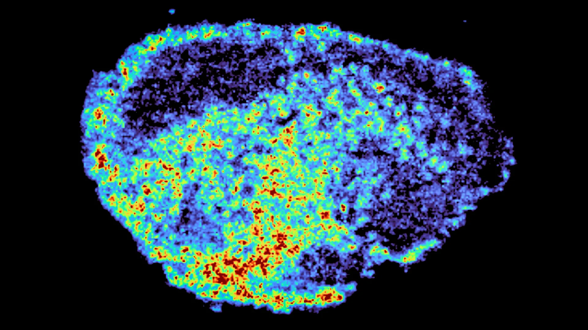
The fundamental mystery of modern oncology lies in the unpredictable nature of therapeutic response: why a drug that eliminates a tumor in one patient may prove entirely ineffective in another with an ostensibly identical diagnosis. This challenge is particularly acute in the treatment of ovarian cancer, where PARP inhibitors have become a cornerstone of therapy but continue to yield inconsistent long-term results. A groundbreaking study published in Nature Communications, led by researchers at the MRC Laboratory of Medical Sciences (LMS) and Imperial College London, has provided a new mechanical explanation for this phenomenon. By utilizing advanced imaging technologies to track drug movement at a cellular level, the team discovered that lysosomes—the cell’s internal "recycling centers"—act as unexpected reservoirs for certain cancer drugs, significantly altering their distribution and effectiveness within the tumor microenvironment.
The Evolution of Ovarian Cancer Therapeutics
Ovarian cancer remains one of the most lethal gynecological malignancies, largely due to its tendency to be diagnosed at an advanced stage when the disease has already spread throughout the peritoneal cavity. For decades, the standard of care was limited to cytoreductive surgery followed by platinum-based chemotherapy. However, the emergence of Poly (ADP-ribose) polymerase (PARP) inhibitors marked a paradigm shift in the mid-2010s. These drugs utilize the principle of "synthetic lethality," specifically targeting cancer cells with pre-existing defects in DNA repair, such as those with BRCA1 or BRCA2 mutations.
While PARP inhibitors like olaparib, rucaparib, and niraparib have significantly extended progression-free survival for many, clinical data has consistently shown a spectrum of resistance. Some patients experience a complete and durable response, while others see their tumors progress within months. Until now, research into this resistance has focused primarily on genetic mutations that allow cancer cells to bypass the PARP blockade. The new research from the MRC LMS suggests that the physical distribution and sequestration of the drug within the cell’s architecture may be just as critical as the genetic makeup of the tumor itself.
Innovative Methodology: The Use of Tumor Explants and Spatial Mapping
To investigate why drug efficacy varies so widely, Dr. Louise Fets and her team moved beyond traditional two-dimensional cell cultures, which often fail to replicate the complex physical barriers of a real human tumor. Instead, they utilized "explants"—thin, live slices of ovarian tumors donated by patients undergoing surgery. These explants were maintained in a laboratory environment that preserved their original architecture, allowing the researchers to observe how drugs penetrate and move through authentic human tissue.
The study employed a sophisticated dual-imaging approach to create a comprehensive map of drug behavior. First, the team used mass spectrometry imaging (MSI), a technique that allows for the visualization of specific molecules across a tissue sample without the need for radioactive labeling. This provided a high-resolution "heat map" of where the PARP inhibitors were accumulating. Simultaneously, the researchers applied spatial transcriptomics, an emerging technology that measures gene activity in specific locations within a tissue slice. By overlaying the drug maps with the gene activity maps, the team could directly compare how cells in "high-drug" areas responded versus those in "low-drug" areas within the same patient sample.
The Discovery of Lysosomal Sequestration
The most striking finding of the study was the uneven distribution of drugs at the single-cell level. The researchers observed that certain PARP inhibitors were not spreading uniformly throughout the cytoplasm to reach their intended target—the DNA in the nucleus. Instead, the drugs were being pulled into lysosomes.
Lysosomes are acidic organelles responsible for breaking down cellular waste. The study revealed that rucaparib and niraparib, due to their specific chemical structures as "cationic amphiphilic drugs," are prone to "lysosomal trapping." Because the interior of a lysosome is significantly more acidic than the surrounding cytoplasm, these drug molecules become chemically altered once they enter, preventing them from diffusing back out easily.
This sequestration creates a "reservoir effect." While it might seem counterintuitive, this trapping can actually be beneficial in some contexts. The lysosomes hold onto the drug and release it slowly over time, potentially providing a sustained therapeutic effect even after the initial dose has cleared the bloodstream. However, if the drug remains trapped too securely or is concentrated in the wrong cells, it leaves other areas of the tumor under-treated, providing an opportunity for cancer cells to survive and develop resistance.
Comparative Analysis of PARP Inhibitors
The research highlighted significant differences between the leading drugs in the PARP inhibitor class. While rucaparib and niraparib showed high levels of lysosomal accumulation, olaparib—the first PARP inhibitor to receive FDA approval—did not. This distinction is critical for clinicians when deciding which therapy to prescribe.
"We were surprised to see large variability in drug accumulation at the single-cell level," noted Dr. Carmen Ramirez Moncayo, the study’s first author. "This variability was driven by the build-up of a drug in lysosomes, which are acting as reservoirs, increasing the exposure of cancer cells to drugs by storing and releasing the drug when needed."
The data suggests that the chemical properties of the drug itself interact with the unique "lysosomal load" of a patient’s tumor. Some tumors may have a higher density of lysosomes or a more acidic internal environment, which would intensify the trapping effect. This adds a layer of complexity to personalized medicine: doctors may eventually need to screen not just for genetic mutations, but for the "physical landscape" of the tumor cells to predict which drug will be most effective.
Supporting Data and Clinical Context
Current clinical statistics underscore the urgency of this research. While approximately 70% of ovarian cancer patients initially respond to platinum-based therapies and subsequent PARP inhibitors, the majority will eventually relapse. Five-year survival rates for advanced ovarian cancer hover around 30% to 40%, a figure that has seen only incremental improvements despite the introduction of targeted therapies.
The findings from the MRC LMS study provide a plausible explanation for "pharmacokinetic resistance," where the drug fails not because the cancer is immune to its mechanism, but because the drug simply cannot reach its target in sufficient concentrations. By identifying lysosomes as the primary site of drug sequestration, the research opens the door to new combination therapies. For instance, drugs that alter lysosomal pH or inhibit lysosomal function could potentially be used alongside PARP inhibitors to "unlock" the trapped medication and ensure a more uniform distribution throughout the tumor.
Expert Reactions and Future Implications
The oncology community has reacted with cautious optimism to these findings. Dr. Zoe Hall, a senior author of the study and Associate Professor at Imperial College London, emphasized the novelty of the spatial mapping approach. "Through the spatial mapping of drug molecules, we could pinpoint regions of high and low drug and compare gene expression from the same tissue slice. This allows us to see exactly how the tumor is fighting back in real-time."
Dr. Louise Fets, Head of the LMS’ Drug Transport and Tumour Metabolism Group, views this as a stepping stone toward a more refined version of precision medicine. "Eventually, we hope to be able to study the molecular signature of a patient’s tumor to help tailor therapeutic approaches in a more personalized way," she stated.
The implications of this research extend beyond ovarian cancer. PARP inhibitors are currently used or being tested for breast, prostate, and pancreatic cancers. If lysosomal trapping is a universal feature of these drugs across different tissue types, the discovery could lead to a wholesale re-evaluation of dosing strategies and drug design for multiple forms of the disease.
Chronology of Research and Funding
The study represents several years of interdisciplinary collaboration, combining expertise in pharmacology, oncology, and advanced imaging. The timeline of the research involved:
- Initial Observation: Identifying that drug concentration in the blood does not always correlate with tumor shrinkage.
- Model Development: Perfecting the use of patient-derived tumor explants to maintain tissue viability outside the body.
- Technological Integration: Combining Mass Spectrometry Imaging with Spatial Transcriptomics—a feat of data integration that allowed for cell-by-cell analysis.
- Data Validation: Testing multiple PARP inhibitors to identify which were susceptible to lysosomal trapping.
The research was made possible through extensive support from major scientific bodies, including the Medical Research Council (MRC) and Cancer Research UK. Additional funding was provided by the Victoria’s Secret Global Fund for Women’s Cancers and the Integrative Toxicology Training Partnership. This diverse backing reflects the study’s importance in both basic biological research and clinical application.
Conclusion: A New Frontier in Precision Oncology
The discovery that lysosomes act as hidden reservoirs for cancer drugs marks a significant shift in our understanding of drug resistance. It suggests that the "geography" of a cell is just as important as its "instruction manual" (the genome). As researchers move forward, the next phase of study will involve animal models to see how the disorganized blood vessels of a living tumor further complicate drug delivery.
By mapping the hidden pockets where drugs accumulate or disappear, scientists are moving closer to a future where cancer treatment is no longer a matter of trial and error. Instead, by analyzing the specific cellular architecture of a patient’s tumor, clinicians may soon be able to prescribe the exact drug, at the exact dose, designed to reach the exact location where it is needed most. This study in Nature Communications is a vital step toward ensuring that the promise of targeted therapy is realized for every patient, regardless of the hidden complexities within their cells.
