
 By George Ongkojoyo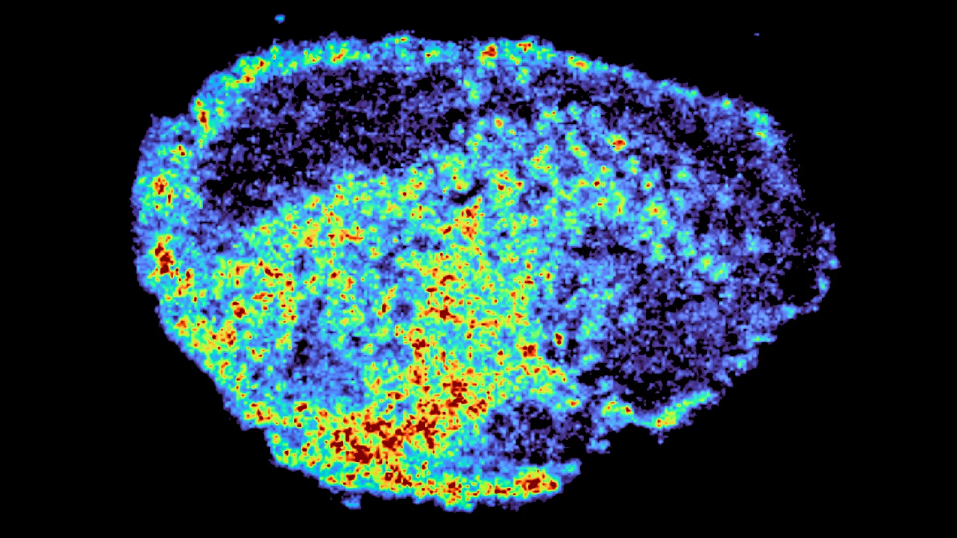
The fundamental paradox of modern oncology lies in the unpredictable nature of therapeutic response: a drug that serves as a life-saving breakthrough for one patient may offer no clinical benefit to another, even when their tumors appear pathologically identical. A seminal study published in the journal Nature Communications has provided a new perspective on this phenomenon by uncovering the microscopic physical barriers and biological "traps" that dictate how drugs move within human tissue. Led by Dr. Louise Fets at the MRC Laboratory of Medical Sciences (LMS), the research team utilized pioneering imaging technologies to track the movement of PARP inhibitors—a vital class of targeted therapies—within ovarian cancer samples. Their findings reveal that the effectiveness of these treatments is deeply influenced by lysosomes, the cellular "recycling centers" that can act as internal reservoirs, sequestering drug molecules and altering their distribution in ways previously unmapped by medical science.
The Challenge of Therapeutic Heterogeneity in Ovarian Cancer
Ovarian cancer remains one of the most lethal gynecological malignancies, often characterized by late-stage diagnosis and a high rate of recurrence. While the advent of Poly (ADP-ribose) polymerase (PARP) inhibitors has revolutionized the treatment landscape—particularly for patients harboring BRCA1 or BRCA2 mutations—the clinical reality is far from uniform. Some patients experience long-term remission, while others see their tumors progress despite high-dose therapy.
The traditional understanding of drug resistance has focused largely on genetic mutations that allow cancer cells to bypass the drug’s mechanism of action. However, the MRC LMS study shifts the focus toward "pharmacological resistance"—the failure of a drug to reach its molecular target at a sufficient concentration. For a PARP inhibitor to be effective, it must penetrate the dense, often chaotic structure of a solid tumor, enter individual cancer cells, and reach the nucleus to inhibit DNA repair enzymes. The researchers discovered that this journey is fraught with obstacles at the sub-cellular level, specifically within the acidic environment of the lysosome.
Methodological Innovation: The Use of Tumor Explants and Spatial Mapping
To observe these dynamics in a setting that mirrors the human body, the research team employed a sophisticated "explant" model. Rather than relying on traditional two-dimensional cell cultures, which fail to capture the complex architecture of a tumor, the scientists used thin slices of live ovarian tumor tissue donated by patients. These samples were maintained in a nutrient-rich environment that preserved the original spatial relationships between cancer cells, connective tissue, and the extracellular matrix.
The study integrated two cutting-edge analytical tools to create a comprehensive map of drug behavior:
- Mass Spectrometry Imaging (MSI): This technique allowed the team to visualize the exact location and concentration of drug molecules across a slice of human tissue. Unlike standard imaging, MSI does not require fluorescent labels, which can sometimes alter the chemical properties of the drug being studied.
- Spatial Transcriptomics: This technology enabled the researchers to measure gene expression within specific regions of the tissue. By overlaying the drug maps from MSI with the genetic maps from spatial transcriptomics, the team could compare the biological "signature" of areas with high drug accumulation against those where the drug failed to penetrate.
This dual-mapping approach revealed a startling level of variability. Even within a single tumor sample treated with a uniform dose, some regions were saturated with the medication while neighboring clusters of cells remained virtually untouched.
The Role of Lysosomes as Hidden Drug Reservoirs
The most significant revelation of the study was the identification of lysosomes as "slow-release reservoirs" for certain PARP inhibitors. Lysosomes are membrane-bound organelles responsible for breaking down waste materials within the cell. Because they maintain a highly acidic internal environment, they can attract and trap "lysosomotropic" molecules—chemicals that are basic (alkaline) in nature.
The research demonstrated that PARP inhibitors like rucaparib and niraparib are particularly prone to being sequestered within these organelles. Once inside the lysosome, the drug molecules become protonated and are unable to easily pass back through the lysosomal membrane into the rest of the cell. This creates internal pockets of high drug concentration.
Dr. Carmen Ramirez Moncayo, the study’s first author and a Postdoctoral Researcher at the LMS, noted the surprise of the team at the sheer scale of this single-cell variability. "The lysosomes act as reservoirs, storing the drug and releasing it gradually over time," Dr. Moncayo explained. This sequestration can be a double-edged sword: while it may increase the duration of exposure for some cells, it can also prevent the drug from reaching the nucleus where it is needed to trigger cell death, potentially allowing some cancer cells to survive and develop further resistance.
Differential Drug Behavior: Why Not All PARP Inhibitors Are Equal
A critical finding for clinicians is that not all drugs in the PARP inhibitor class behave the same way. The study compared three major inhibitors: rucaparib, niraparib, and olaparib. While rucaparib and niraparib showed significant lysosomal accumulation, olaparib—a drug with different chemical properties—distributed more evenly throughout the cell and was not trapped by the lysosomal "recycling centers."
This distinction is vital for the future of personalized oncology. If a patient’s tumor is found to have a high density of lysosomes or a specific pH profile that favors sequestration, a physician might choose a non-lysosomotropic drug like olaparib to ensure more uniform distribution. Conversely, in other scenarios, the "slow-release" effect of rucaparib might be therapeutically advantageous. Understanding these chemical nuances allows for a more tactical approach to drug selection.
Official Perspectives and the Path to Personalized Care
The implications of this research extend beyond the laboratory, offering a roadmap for more precise clinical trials. Dr. Zoe Hall, senior author and Associate Professor at Imperial College London’s Department of Metabolism, Digestion and Reproduction, emphasized the novelty of the study’s direct measurement techniques. "Through the spatial mapping of drug molecules, we could pinpoint regions of high and low drug and compare gene expression from the same tissue slice," Dr. Hall stated. This ability to link drug concentration directly to biological response at a microscopic level is a significant leap forward from traditional blood-concentration monitoring.
Dr. Louise Fets, Head of the LMS’ Drug Transport and Tumour Metabolism Group, expressed hope that this molecular-level understanding will eventually lead to a "molecular signature" for each patient. "By understanding how drugs are taken up into cells, we can understand whether this influences why cancer drugs work for some people and not for others," Dr. Fets said. The ultimate goal is to tailor therapeutic approaches so that the specific physical and chemical characteristics of a patient’s tumor dictate the choice of medication, rather than a "trial and error" method.
Broader Impact and Future Research Directions
The study opens several new avenues for investigation. While the research utilized tumor explants, the team acknowledges that the delivery of drugs in a living patient is even more complex due to the disorganized nature of tumor vasculature. In a clinical setting, poor blood flow can prevent a drug from reaching the tumor at all, adding another layer of difficulty to the distribution issues identified in this study.
Future research phases will involve:
- Animal Models: Testing how lysosomal trapping interacts with the circulatory system in vivo.
- Relapsed Cancer Studies: Investigating whether tumors that have become resistant to treatment show increased lysosomal sequestration as a defense mechanism.
- Drug Design: Developing new iterations of PARP inhibitors that are engineered to either exploit lysosomal storage or bypass it entirely to reach the cell nucleus more efficiently.
The broader scientific community has welcomed these findings as a necessary step toward solving the "black box" of drug distribution. As cancer treatment moves toward increasingly targeted therapies, the physical journey of the molecule—from the IV drip or pill to the microscopic structures within a single cell—is becoming as important as the genetic makeup of the cancer itself.
Acknowledgments and Funding
The research was a collaborative effort supported by a diverse range of prestigious institutions. Funding was provided by the Medical Research Council (MRC) and Cancer Research UK. Additional support came from a PhD studentship via the Integrative Toxicology Training Partnership (administered by the MRC Toxicology Unit) and a Victoria’s Secret Global Fund for Women’s Cancers Career Development Award, provided in partnership with Pelotonia and the American Association for Cancer Research (AACR).
This interdisciplinary support underscores the global importance of the work, as PARP inhibitors are currently being tested for efficacy in breast, prostate, and pancreatic cancers, potentially expanding the impact of these findings to hundreds of thousands of patients worldwide. As the medical community continues to refine its approach to precision medicine, the "hidden reservoirs" of the lysosome will undoubtedly remain a focal point for researchers striving to ensure that every patient receives a therapy that is not only targeted but also accessible to the cells that need it most.
