
 By George Ongkojoyo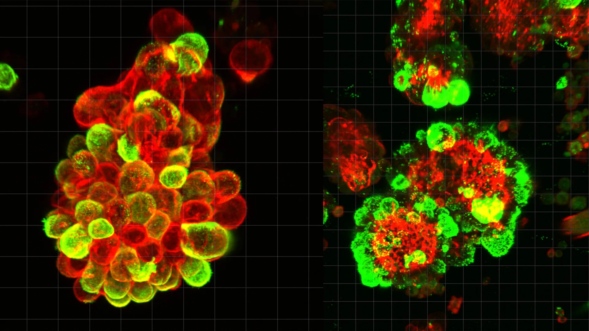
Ovarian cancer has long held a reputation as one of the most formidable challenges in oncology, primarily due to its ability to spread silently and rapidly throughout the abdominal cavity before clinical symptoms emerge. For decades, the biological mechanisms that allow these cancer cells to colonize the peritoneum with such devastating efficiency remained partially obscured. However, a groundbreaking study led by a research team at Nagoya University in Japan has finally unmasked the "secret accomplice" in this process. The research, recently published in the prestigious journal Science Advances, reveals that ovarian cancer cells do not act in isolation; instead, they hijack healthy mesothelial cells—the very cells meant to protect the abdominal lining—to facilitate their invasion and shield themselves from the effects of chemotherapy.
The discovery centers on the formation of "hybrid cell clusters," a sophisticated biological partnership that transforms the abdominal cavity into a highway for malignancy. By understanding how these clusters form and function, researchers believe they have identified a critical vulnerability that could lead to more effective treatments and earlier diagnostic tools for a disease that currently claims more lives than any other gynecological malignancy.
The Mechanism of Co-opted Invasion: How Hybrid Clusters Form
The traditional understanding of cancer metastasis often focuses on the intrinsic mutations of the cancer cell itself. While these mutations are vital, the Nagoya University study highlights the role of the "microenvironment"—the surrounding cellular landscape. In the case of ovarian cancer, the primary environment is the peritoneal cavity, which is lined by a thin layer of mesothelial cells.
Under normal physiological conditions, mesothelial cells provide a smooth, protective, and non-adhesive surface that allows organs to slide against one another. However, when ovarian cancer cells detach from the primary tumor in the ovaries or fallopian tubes, they enter the peritoneal fluid (ascites). The researchers discovered that these floating cancer cells actively recruit mesothelial cells that have shed from the abdominal lining.
Using high-resolution microscopy and single-cell RNA sequencing, the team observed that approximately 60% of the cancer cell spheres found in the abdominal fluid of patients were not pure cancer. Instead, they were hybrid spheroids consisting of both malignant cells and recruited mesothelial cells. This recruitment is not accidental but is driven by a specific biochemical "handshake." The cancer cells secrete a potent signaling molecule known as Transforming Growth Factor-beta 1 (TGF-β1). This protein acts as a reprogramming agent, fundamentally altering the behavior and structure of the healthy mesothelial cells.
Invadopodia: The Biological Drills of the Abdomen
Once the mesothelial cells are integrated into the hybrid cluster and activated by TGF-β1, they undergo a transformation that makes them more aggressive than the cancer cells themselves. The study found that these "hijacked" mesothelial cells begin to develop invadopodia—specialized, spike-like protrusions rich in actin filaments and digestive enzymes.
These invadopodia act as biological drills. When a hybrid cluster makes contact with a new organ—such as the liver, the omentum, or the intestinal wall—the mesothelial cells lead the way. They use their invadopodia to puncture the protective lining of the organ, creating physical openings. The cancer cells, remaining relatively passive, simply follow these pathways to establish new metastatic colonies.
Dr. Kaname Uno, the study’s lead author and a Visiting Researcher at Nagoya University’s Graduate School of Medicine, noted that this strategy allows cancer cells to conserve energy and avoid the need for complex genetic mutations usually required for tissue invasion. "The cancer cells manipulate mesothelial cells to do the heavy lifting of tissue invasion," Dr. Uno explained. "They undergo minimal genetic and molecular changes themselves, essentially migrating through the breaches created by their recruited accomplices."
A Departure from Conventional Metastasis
The spread of ovarian cancer represents a significant departure from the metastatic pathways utilized by other common cancers, such as breast, lung, or prostate cancer. In those diseases, the primary route of spread is hematogenous—meaning cancer cells must penetrate blood vessels, survive the turbulent environment of the bloodstream, and then exit the vessels at a distant site. Because blood follows a predictable circuit, clinicians can often monitor these cancers through blood-based "liquid biopsies" that detect circulating tumor cells (CTCs) or cell-free DNA.
Ovarian cancer, by contrast, utilizes "peritoneal dissemination." The cancer cells bypass the bloodstream entirely, instead drifting through the peritoneal fluid. This fluid is in a state of constant, unpredictable flux, driven by the movement of the diaphragm during breathing and the peristaltic motion of the intestines. This lack of a fixed route makes the spread of ovarian cancer exceptionally difficult to track or predict. The Nagoya University study provides the first comprehensive look at what happens during this "floating phase," proving that it is not merely a passive transit period but an active phase of assembly and preparation for invasion.
The Shield of Chemoresistance
Beyond facilitating invasion, the hybrid cell clusters offer a second, equally dangerous advantage: enhanced resistance to chemotherapy. Standard treatments for ovarian cancer typically involve platinum-based drugs designed to kill rapidly dividing cells. However, when cancer cells are tucked inside a hybrid sphere with mesothelial cells, they become significantly harder to eradicate.
The study’s data suggests that the mesothelial cells provide a physical and biochemical shield. By forming a compact, three-dimensional structure, the hybrid clusters limit the penetration of chemotherapy drugs into the core of the sphere. Furthermore, the interaction between the two cell types may trigger "survival signaling" that prevents the cancer cells from undergoing apoptosis (programmed cell death) even when exposed to toxic agents. This explains why many ovarian cancer patients initially respond well to chemotherapy, only to experience a recurrence of the disease that is no longer sensitive to standard drugs.
From the Clinic to the Lab: The Motivation of Dr. Kaname Uno
The impetus for this research was deeply personal for the lead scientist. Before entering the world of molecular research, Dr. Kaname Uno spent eight years as a practicing gynecologist. During his time in the clinic, he witnessed the limitations of current diagnostic technology firsthand.
One particular case stayed with him: a patient who had undergone a routine gynecological screening and received a clean bill of health. Only three months later, she returned with severe abdominal symptoms and was diagnosed with Stage IV ovarian cancer. The disease had spread so rapidly and extensively that it had evaded detection just 90 days prior.
"The inability of our current tools to save patients like her was a profound motivation," Dr. Uno said. "I needed to understand the biological ‘why’ behind this speed. Why does this cancer move faster than we can detect it?" His transition from the operating room to the laboratory was driven by the realization that until the mechanics of peritoneal spread were understood, survival rates for advanced ovarian cancer would remain stagnantly low.
Statistical Context and the "Silent Killer"
The urgency of this research is underscored by the global statistics surrounding ovarian cancer. According to the World Health Organization (WHO), more than 300,000 new cases are diagnosed annually, with a high mortality-to-incidence ratio. Because the symptoms—bloating, pelvic pain, and feeling full quickly—are often vague and easily confused with digestive issues, roughly 75% of patients are diagnosed at Stage III or IV.
At these advanced stages, the five-year survival rate drops significantly compared to cancers caught in Stage I. The Nagoya University study provides a scientific explanation for why the "silent killer" is so effective: by the time a patient feels bloated, the hybrid clusters have already used their invadopodia to anchor themselves across the abdominal cavity, creating a multi-focal disease that is difficult to surgically resect or chemically treat.
Implications for Future Therapy and Diagnostics
The identification of the TGF-β1 signaling pathway and the role of mesothelial invadopodia opens several new avenues for clinical intervention.
- Targeting the Partnership: Current chemotherapy is designed to kill cancer cells. Future therapies could be "combination" treatments that also target the mesothelial cells or block the TGF-β1 signal that allows the hybrid clusters to form. By preventing the "partnership," doctors could potentially keep the cancer cells in a floating, non-invasive state that is more vulnerable to treatment.
- Inhibiting Invadopodia: Drugs that inhibit the formation of invadopodia are already being explored in other areas of cancer research. Applying these to ovarian cancer could stop the "drilling" process, preventing the cancer from anchoring to vital organs.
- New Diagnostic Markers: Rather than relying solely on the CA-125 blood test—which can be unreliable—doctors might one day analyze peritoneal fluid for the presence of these specific hybrid cell clusters. Detecting these clusters in the early stages of formation could provide a much more accurate picture of a patient’s prognosis and how aggressively the cancer is likely to spread.
- Refined Liquid Biopsies: While blood tests have limitations in ovarian cancer, "peritoneal liquid biopsies" (sampling the abdominal fluid) could become a standard way to monitor treatment response. If a treatment successfully breaks apart the hybrid clusters, it could be a sign that the cancer’s invasive potential has been neutralized.
Conclusion: A New Paradigm in Ovarian Cancer Research
The research from Nagoya University marks a paradigm shift in how scientists view ovarian cancer metastasis. By shifting the focus from the cancer cell alone to the "hybrid partnership" it forms with the mesothelium, the study provides a roadmap for the next generation of oncological care.
For years, the speed of ovarian cancer was a mystery that left doctors one step behind. By uncovering the role of the mesothelial "accomplice" and the mechanical "drills" they use to invade tissue, researchers have finally caught up. The challenge now lies in translating these laboratory insights into bedside treatments that can break the cycle of rapid spread and chemoresistance, offering hope to thousands of women who face this devastating diagnosis.
