
 By Billy Candra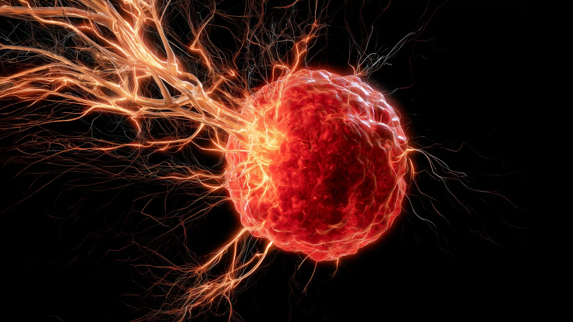
New research emanating from the University of Oklahoma (OU) has cast significant light on a complex interplay within the tumor microenvironment, revealing how a particularly aggressive form of breast cancer, triple-negative breast cancer (TNBC), can strategically manipulate the body’s immune system. This manipulation leads to the recruitment of nerves into tumor masses, establishing conditions that appear to facilitate cancer progression and potentially contribute to treatment resistance. The groundbreaking findings, recently published in the esteemed scientific journal Cell Death & Differentiation, address a long-standing question in oncology regarding the precise mechanisms by which nerves infiltrate solid tumors, offering a novel avenue for therapeutic intervention.
For decades, oncologists and pathologists have observed the presence of extensive nerve networks within various solid tumors. However, the exact process by which these nerves are drawn into the cancerous tissue, rather than simply being displaced by it, has remained largely enigmatic. This latest study from the University of Oklahoma provides a critical explanation for this phenomenon, specifically in the context of triple-negative breast cancer, a subtype notorious for its challenging prognosis and limited treatment options compared to other breast cancer types.
Understanding Triple-Negative Breast Cancer: An Aggressive Challenge
Triple-negative breast cancer represents approximately 10-15% of all diagnosed breast cancers, yet it accounts for a disproportionately higher percentage of breast cancer deaths. Its "triple-negative" designation refers to the absence of three common receptors that fuel most breast cancers: estrogen receptors (ER), progesterone receptors (PR), and human epidermal growth factor receptor 2 (HER2). The lack of these receptors means that TNBC does not respond to hormone therapy (which targets ER/PR) or HER2-targeted therapies (like Herceptin). Consequently, treatment options are primarily limited to chemotherapy, surgery, and radiation, often with less favorable outcomes and a higher risk of recurrence and metastasis. The aggressive nature of TNBC, its tendency to affect younger women, and its poorer survival rates underscore the urgent need for innovative research to uncover new therapeutic targets and strategies. The OU research offers a potential paradigm shift in understanding and combating this formidable disease.
The Immune System’s Unwitting Complicity: Macrophages as Facilitators
The core of the OU team’s discovery lies in identifying the role of macrophages, a type of immune cell traditionally recognized for its protective functions. Macrophages are vital components of the innate immune system, serving as phagocytes that engulf cellular debris, pathogens, and foreign substances. They are also crucial for tissue repair and inflammation resolution. In a healthy physiological context, macrophages are indispensable for maintaining tissue homeostasis and defending against threats.
However, the OU researchers found that in the context of triple-negative breast cancer, tumors cunningly co-opt these immune cells. The cancer cells attract macrophages into the tumor microenvironment, where these immune cells undergo a transformation, often referred to as polarization, becoming "tumor-associated macrophages" (TAMs). Once integrated into the tumor, these TAMs, instead of mounting an anti-tumor response, begin to secrete a specific protein: brain-derived neurotrophic factor (BDNF).
BDNF: A Double-Edged Sword in Cancer Biology
BDNF is a neurotrophin, a family of proteins known for their crucial roles in the development, maintenance, and survival of neurons in the central and peripheral nervous systems. It is most widely recognized for supporting the growth, differentiation, and survival of nerve cells in the brain, playing a vital role in learning, memory, and cognitive function. Its discovery in the mid-1980s marked a significant advance in neurobiology, leading to extensive research into its therapeutic potential for neurodegenerative diseases.
Yet, in the context of breast cancer, the OU study reveals a darker side to BDNF’s biological signaling. The research team demonstrated that TNBC tumors exploit this very same biological signal. By prompting tumor-associated macrophages to release BDNF, the cancer effectively creates a chemical gradient that encourages nearby nerves to grow toward and directly into the tumor mass. This process of tumor innervation, driven by BDNF, is now understood to be a significant contributor to cancer progression and, alarmingly, resistance to current treatments.
Dr. Maureen Cox, Ph.D., an assistant professor in the Department of Microbiology and Immunology at the OU College of Medicine and a research member of OU Health Stephenson Cancer Center, articulated the paradoxical role of macrophages in this scenario. "Macrophages are the critical source for drawing nerves into the tumor," Dr. Cox stated. "Although macrophages typically play a positive role in the body, they are facilitating a negative function in this scenario of breast cancer." Her statement underscores the complexity of the tumor microenvironment, where the very cells designed to protect the body can be reprogrammed to inadvertently support cancer growth.
Chronology of Discovery and the Evolution of Understanding Tumor Innervation
The recognition of nerves within tumors is not entirely new. Early histological studies in the 19th and 20th centuries occasionally noted nerve fibers amidst cancerous cells. However, for a long time, these observations were largely considered coincidental, with nerves simply being trapped or displaced by the expanding tumor mass. The active recruitment and functional integration of nerves into tumors—a concept now termed "tumor innervation"—began to gain serious scientific attention in the late 20th and early 21st centuries.
Initial studies focused on the sympathetic and parasympathetic nervous systems, hypothesizing that neurotransmitters might influence cancer cell behavior. Subsequent research began to uncover specific growth factors released by tumors that could attract nerves, similar to how tumors recruit blood vessels (angiogenesis). The OU study marks a significant milestone in this chronological understanding, pinpointing a precise cellular orchestrator (macrophages) and a specific molecular signal (BDNF) responsible for this innervation in an aggressive cancer type. This level of mechanistic detail represents a substantial leap forward, moving from mere observation to functional explanation.
Blocking the Signal: A Promising Therapeutic Avenue
The discovery of BDNF’s role immediately suggested a potential therapeutic strategy: interrupting this signaling pathway. The OU team translated this hypothesis into preclinical studies, testing their approach in mouse models of triple-negative breast cancer. They employed a drug known to block BDNF signaling and observed compelling results. In mice treated with this inhibitor, the growth of nerves into the tumors was significantly curtailed. Crucially, this reduction in nerve innervation correlated with a substantial decrease in overall tumor growth.
This finding is particularly exciting because the drug used in their experiments is already "on the market," as Dr. Cox noted. The concept of drug repurposing, where an existing drug approved for one condition is found to be effective for another, holds immense promise in oncology. Repurposed drugs have a known safety profile, established dosing regimens, and can bypass many of the lengthy and expensive stages of early drug development. This could drastically accelerate the timeline for bringing a new treatment strategy to patients with TNBC, potentially offering a more immediate impact than developing an entirely novel compound.
"It looks really promising that we can use this drug, which is already on the market, to target BDNF," Dr. Cox emphasized, highlighting the clinical translation potential. Beyond directly inhibiting tumor growth, the researchers hypothesize a broader benefit. "We believe that the nerves are immunosuppressive, so if we can stop the nerves from growing in the first place, maybe we can boost the immune response to help fight the cancer." This suggests a dual advantage: direct anti-tumor effects and an enhanced ability for the body’s own immune system to recognize and attack cancer cells, a critical goal in modern cancer therapy, particularly immunotherapy.
Evidence from Human Patients: Validating the Mechanism
To ascertain the clinical relevance of their findings from mouse models, the OU researchers extended their investigation to human data. They analyzed samples and clinical information from patients diagnosed with triple-negative breast cancer. Their analysis revealed a compelling correlation: tumors from patients with higher levels of both macrophages and BDNF were linked with poorer overall survival outcomes. This crucial piece of evidence provides robust support for the translational potential of their work, indicating that the intricate mechanism observed in preclinical models is likely highly relevant to human disease progression and patient prognosis. Such validation is vital for advancing research from the laboratory to the clinic.
Broader Impact and Future Directions: Unraveling the Nerve-Cancer Nexus
The OU research opens numerous avenues for future investigation. While the study clearly demonstrates how nerves are drawn into tumors, the exact mechanisms by which these nerves then contribute to tumor growth remain an area of active exploration. Dr. Cox and her team are now dedicated to better understanding this "how" and "why."
Several hypotheses are being explored. One line of evidence suggests that nerves might stimulate the formation of new blood vessels, a process known as angiogenesis. Tumors are highly dependent on a robust blood supply to deliver oxygen and nutrients for their rapid growth and to remove metabolic waste products. If nerves facilitate angiogenesis, blocking innervation could indirectly starve the tumor.
Another compelling hypothesis posits that cancer cells may utilize nerve fibers as "highways" for metastasis. As cancer cells become more invasive, they might migrate along these established neural pathways, using them to exit the primary tumor site and spread to distant organs. This concept, known as "perineural invasion," is a known prognostic factor in certain cancers, particularly those of the head and neck, and if applicable to breast cancer, blocking innervation could potentially impede metastatic spread.
The researchers also plan to test their intervention strategy—blocking BDNF signaling—in other aggressive cancers that are difficult to treat. High-grade ovarian cancer is a prime candidate, given its similar characteristics of aggressive growth, late diagnosis, and often poor prognosis. If the mechanism of nerve recruitment via macrophages and BDNF is conserved across different aggressive cancer types, the therapeutic implications could be far-reaching.
Ultimately, the overarching goal of Dr. Cox’s team, as she articulates, is profoundly patient-centric: "Ultimately, we want to turn the anti-tumor immunity back on in cancer patients so their own immune systems can reject the tumors." This vision aligns perfectly with the current frontier of cancer research, which increasingly focuses on leveraging the body’s own immune defenses to combat malignancies. By disrupting the tumor’s ability to manipulate its microenvironment and evade immune surveillance, this research aims to unlock the full potential of anti-tumor immunity.
Collaborative Science and Funding Support
This pioneering research underscores the collaborative nature of modern scientific discovery and the critical role of sustained funding. The project received substantial support from multiple prestigious institutions, including the National Institute of General Medical Sciences of the NIH (under award numbers P20GM103447 and P20GM103639). Further crucial backing was provided by Oklahoma’s Tobacco Settlement Endowment Trust (TSET), which serves as a primary funder for the Stephenson Cancer Center and the TSET Health Promotion Research Center at the University of Oklahoma. Additional support came from the Oklahoma Shared Clinical and Translational Resources, through an Institutional Development Award from the National Institute of General Medical Sciences (grant no. U54GM104938). This broad base of funding highlights the recognized importance and potential impact of the research on public health.
In conclusion, the University of Oklahoma’s latest research represents a significant advance in understanding the complex dynamics within the tumor microenvironment of triple-negative breast cancer. By elucidating how aggressive cancers hijack immune cells to attract nerves through BDNF signaling, the study not only solves a long-standing mystery but also unveils a novel, targetable pathway for therapeutic intervention. The promise of repurposing an existing drug to disrupt this pathway offers a beacon of hope for patients battling this challenging disease, potentially leading to more effective treatments and improved outcomes by both directly impeding tumor growth and enhancing the body’s innate immune response against cancer.
