
 By George Ongkojoyo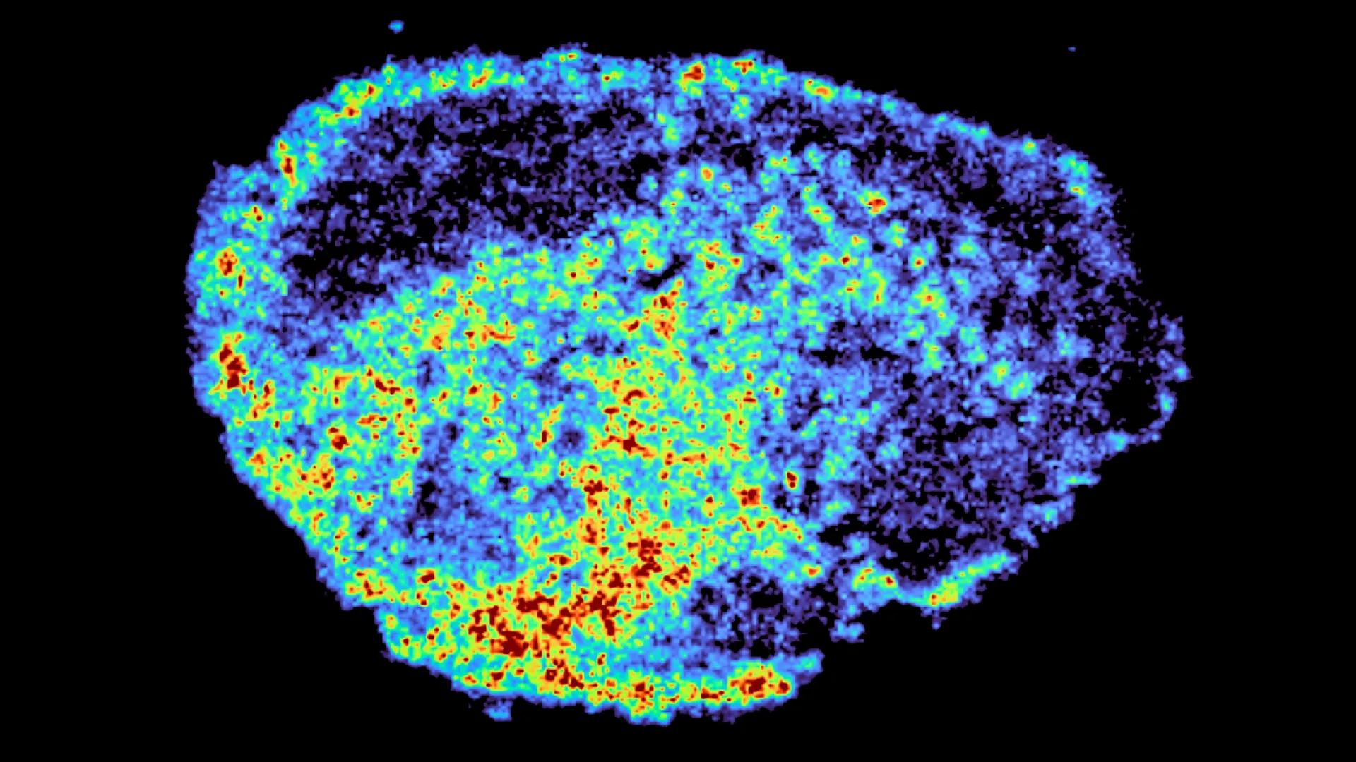
A groundbreaking study published in the journal Nature Communications has unveiled a critical mechanism explaining why highly touted cancer therapies often yield inconsistent results across different patient populations. Led by researchers at the Medical Research Council (MRC) Laboratory of Medical Sciences (LMS), the study provides a high-resolution look at the internal journey of PARP inhibitors within ovarian tumor tissues. By utilizing sophisticated imaging technologies, the team discovered that these drugs frequently become trapped within lysosomes—small, acidic organelles often described as the cell’s "recycling centers." This sequestration creates internal reservoirs that dictate the timing and intensity of drug exposure, offering a new perspective on the persistent challenge of drug resistance and therapeutic failure in oncology.
The research, headed by Dr. Louise Fets, focuses on a class of drugs known as Poly (ADP-ribose) polymerase (PARP) inhibitors. These agents have revolutionized the treatment landscape for ovarian, breast, and prostate cancers, particularly for patients harboring BRCA mutations. However, despite their clinical success, a significant portion of patients either fail to respond to the treatment or develop resistance shortly after the initiation of therapy. The findings from the MRC LMS team suggest that the uneven distribution of these drugs at a cellular level, driven by lysosomal activity, may be a primary culprit behind these clinical discrepancies.
The Pharmacokinetic Challenge in Modern Oncology
For a targeted cancer therapy to be effective, it must not only reach the tumor site via the systemic circulation but also penetrate the dense architecture of the tumor and accumulate within the malignant cells at a concentration sufficient to trigger programmed cell death. Historically, pharmacologists have relied on blood plasma levels to estimate drug efficacy. However, plasma concentrations rarely reflect the true concentration of a drug at its intracellular target.
In the case of PARP inhibitors, the drugs must reach the nucleus of the cancer cell to inhibit DNA repair enzymes. If the drug is diverted or "caged" within other cellular compartments before reaching the nucleus, its therapeutic potential is severely diminished. The study by Dr. Fets and her colleagues highlights that the tumor microenvironment is far from a uniform sponge; it is a complex, heterogeneous landscape where drug molecules are subjected to various biological barriers and diversions.
To investigate this phenomenon, the researchers employed "explants"—thin slices of human ovarian tumor tissue obtained directly from patients and maintained in a viable state within a laboratory setting. This method allowed the scientists to bypass the limitations of traditional 2D cell cultures, which often fail to replicate the three-dimensional structural complexity and cellular diversity of a real human tumor.
Advanced Mapping: Mass Spectrometry and Spatial Transcriptomics
The technical core of the study involved a combination of mass spectrometry imaging (MSI) and spatial transcriptomics. Mass spectrometry imaging allowed the team to create high-resolution chemical maps of the tumor slices, pinpointing exactly where the drug molecules were accumulating. Unlike traditional imaging, which might require fluorescent tagging—a process that can alter the chemical properties of the drug—MSI tracks the drug in its native state.
By pairing these chemical maps with spatial transcriptomics, the researchers could overlay drug distribution data with gene expression profiles. This allowed them to see how cells in "high-drug" regions differed from those in "low-drug" regions within the same patient sample. The results were startling: even within a single tumor slice treated with a uniform dose, drug concentration varied wildly from one cell cluster to the next.
Dr. Zoe Hall, a senior author of the study and Associate Professor at Imperial College London’s Department of Metabolism, Digestion and Reproduction, emphasized the novelty of this dual-mapping approach. "Through the spatial mapping of drug molecules, we could pinpoint regions of high and low drug and compare gene expression from the same tissue slice. This allowed us to understand the biological state of the cells in relation to the actual amount of therapy they received," Dr. Hall explained.
The Role of Lysosomes as Hidden Drug Reservoirs
The most significant discovery of the study was the identification of lysosomes as the primary drivers of drug heterogeneity. The researchers found that certain PARP inhibitors, specifically rucaparib and niraparib, are lysosomotropic, meaning they are chemically drawn into the acidic environment of the lysosome. Once inside, these drugs become protonated and trapped, effectively sequestered away from their intended targets in the cell nucleus.
Interestingly, these lysosomes do not merely act as "trash cans" that neutralize the drug. Instead, they function as slow-release reservoirs. As the concentration of the drug outside the lysosome drops, the trapped drug is gradually released back into the cytoplasm. While this can extend the duration of drug exposure for some cells, it also means that the immediate "hit" required to kill a fast-growing cancer cell may be insufficient.
The study also noted a significant difference between various PARP inhibitors. While rucaparib and niraparib showed high levels of lysosomal sequestration, olaparib—another widely used PARP inhibitor—did not exhibit the same behavior. This distinction is crucial for clinicians, as it suggests that the chemical structure of a specific drug within the same class can radically alter its distribution pattern and, consequently, its clinical performance in different patients.
"We were surprised to see large variability in drug accumulation at the single-cell level," said Dr. Carmen Ramirez Moncayo, the study’s first author. "This variability was driven by the build-up of a drug in lysosomes, which act as reservoirs, increasing the exposure of cancer cells to drugs by storing and releasing the drug when needed. However, if too much is stored and not enough reaches the nucleus, the cell survives."
Chronology of PARP Inhibitor Development and Clinical Use
The discovery of the lysosomal "trap" comes at a pivotal time in the history of PARP inhibitors. The timeline of these drugs reflects a rapid evolution in cancer care:
- 2005: Research published in Nature identifies the concept of "synthetic lethality," showing that PARP inhibitors could specifically kill cells with BRCA1 or BRCA2 mutations.
- 2014: The FDA grants the first accelerated approval for olaparib (Lynparza) for advanced ovarian cancer.
- 2017: Niraparib and rucaparib receive FDA approval, expanding the toolkit for oncologists.
- 2018-2022: Clinical trials expand the use of PARP inhibitors to first-line maintenance therapy and to other cancers, including breast, pancreatic, and prostate.
- 2024: The MRC LMS study identifies lysosomal sequestration as a major factor in drug distribution heterogeneity, providing a molecular basis for varying patient responses.
This timeline illustrates a shift from broad application to a deeper, more mechanistic understanding of how these drugs interact with human biology. The MRC LMS study represents the next phase of this evolution: moving toward "precision pharmacokinetics."
Implications for Personalized Medicine and Future Research
The findings have profound implications for the future of personalized oncology. Currently, cancer treatment is often a process of trial and error, where patients are moved from one line of therapy to another as resistance develops. By understanding the molecular signature of a patient’s tumor—including its lysosomal density and activity—doctors may eventually be able to predict which PARP inhibitor will be most effective.
Dr. Louise Fets, Head of the LMS Drug Transport and Tumour Metabolism Group, noted that this research opens the door to tailoring therapeutic approaches. "Eventually, we hope to be able to study the molecular signature of a patient’s tumor to help tailor therapeutic approaches in a more personalized way. If we can identify which tumors will sequester certain drugs, we can choose an alternative medication that bypasses those compartments," she stated.
The study also suggests a potential new strategy for overcoming drug resistance: the use of lysosomal modulators. If a drug like rucaparib is being trapped in lysosomes, combining it with a second agent that alters lysosomal pH or permeability could "unlock" the drug, forcing it into the nucleus where it can perform its function.
Future Directions and Clinical Integration
While the study provides a detailed map of drug behavior in "explant" models, the researchers acknowledge that the journey of a drug in a living patient is even more complex. In a clinical setting, the drug must navigate a disorganized network of tumor blood vessels, which are often leaky and inefficient. This "macro-scale" distribution problem likely compounds the "micro-scale" lysosomal problem identified in the study.
Future research phases will involve animal models to observe how systemic drug delivery interacts with tumor structure and lysosomal storage in real-time. Additionally, the team plans to study larger patient cohorts, particularly those with relapsed cancers, to determine if lysosomal sequestration increases as tumors evolve and become more resistant to treatment.
The research was a collaborative effort supported by a diverse array of funding bodies, including the Medical Research Council, Cancer Research UK, and the Victoria’s Secret Global Fund for Women’s Cancers. This interdisciplinary support underscores the importance of the findings for the broader medical community.
As oncology moves further into the era of precision medicine, the ability to visualize and quantify drug distribution at the cellular level will become an essential tool. The work of the MRC LMS team provides a vital piece of the puzzle, turning the "black box" of drug delivery into a transparent map that could lead to more effective, more durable treatments for thousands of patients worldwide.
