
 By George Ongkojoyo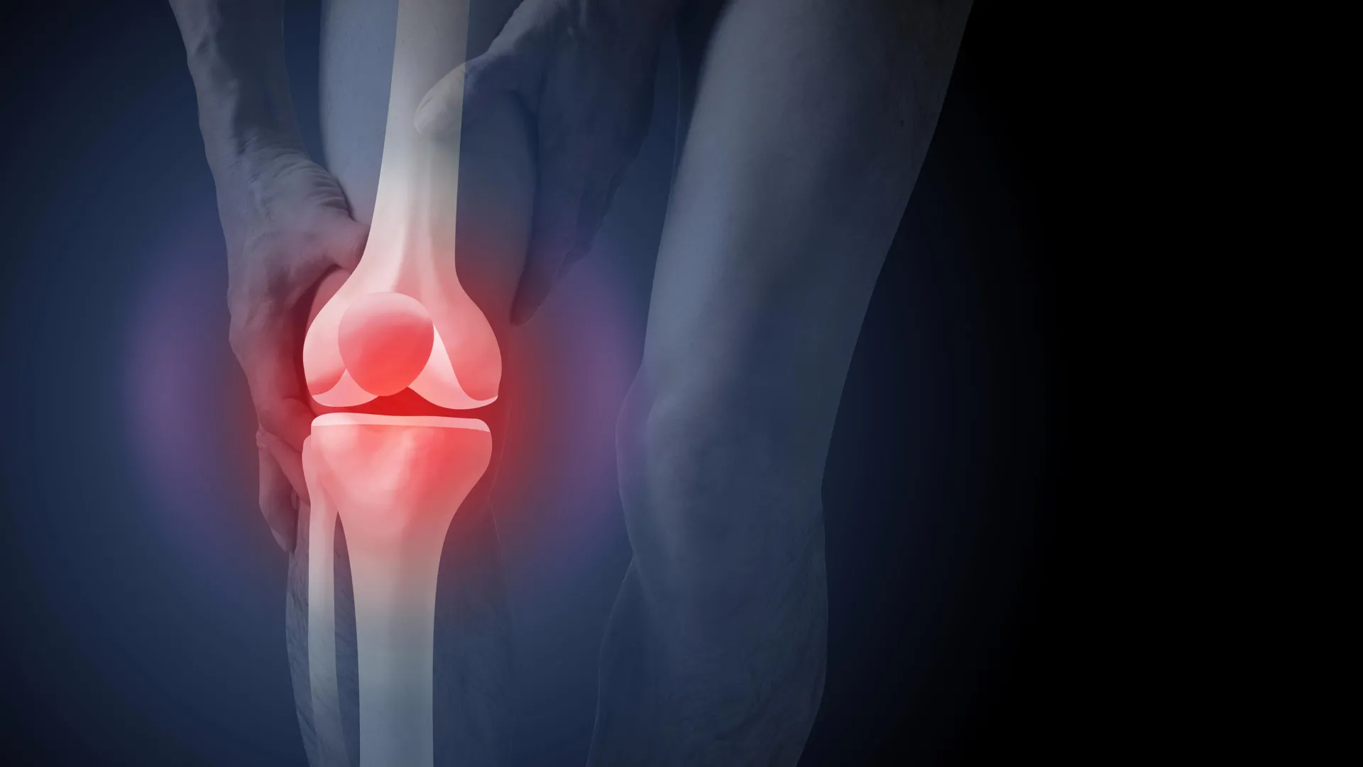
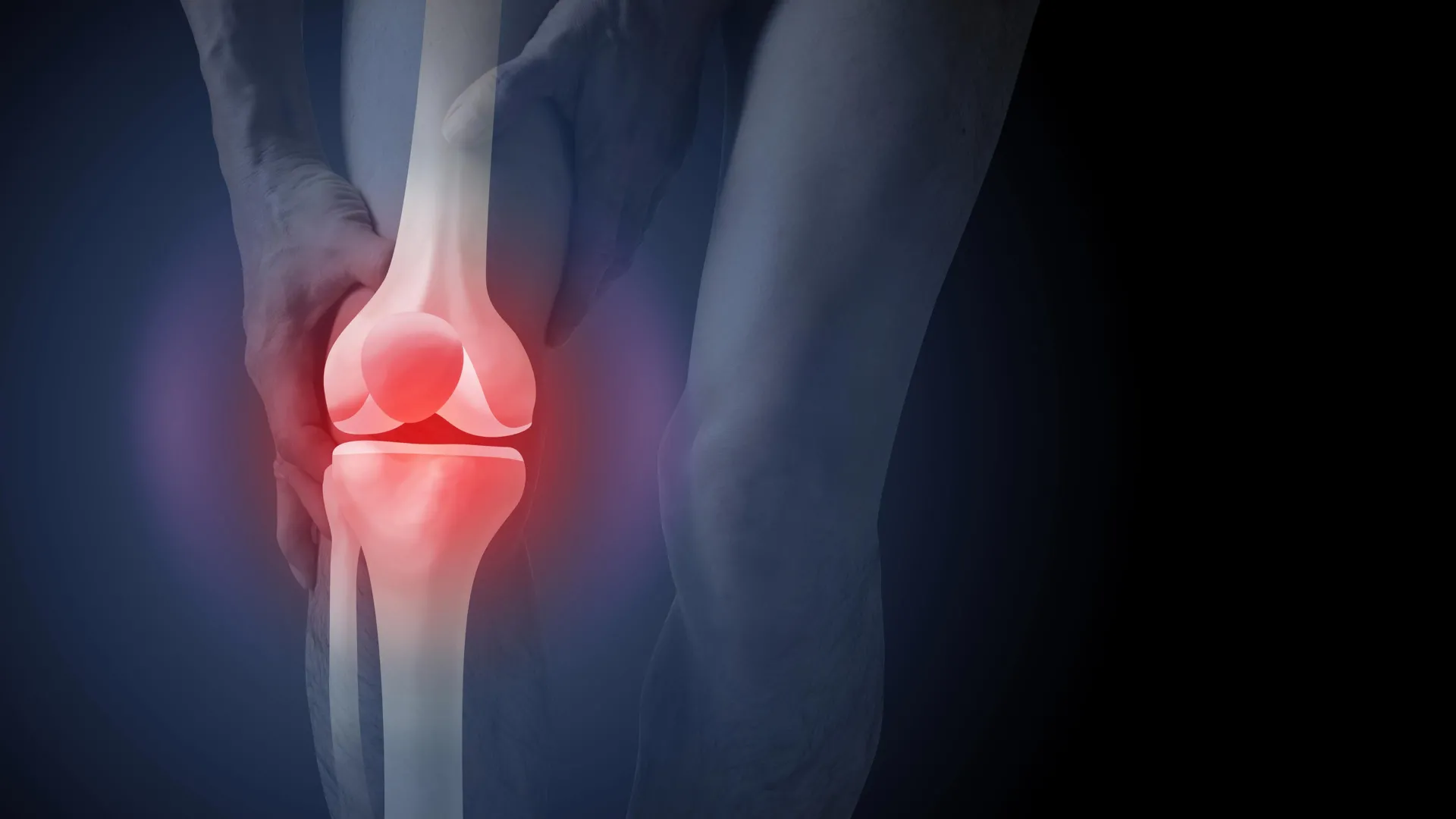
The widespread practice of utilizing routine X-rays to diagnose knee osteoarthritis is increasingly coming under scrutiny as new research highlights significant psychological and economic drawbacks to this approach. Despite clinical guidelines recommending against the use of imaging for initial diagnosis, nearly half of all new patients presenting with knee pain to general practitioners in Australia are referred for X-rays. This diagnostic trend not only costs the national healthcare system over A$104 million annually but also appears to negatively influence patient perceptions of their condition, often steering them toward invasive and potentially unnecessary surgical interventions. A recent study published in PLOS Medicine has provided empirical evidence that viewing X-ray images can lead patients to believe their joints are "worn out," fostering a fear of physical activity and an increased desire for total knee replacement surgery, even when non-surgical alternatives remain the gold standard for care.
Knee osteoarthritis is a chronic condition characterized by changes within the entire joint environment, including the degradation of cartilage, remodeling of underlying bone, and inflammation of the ligaments and muscles. It is a leading cause of disability globally, particularly among older adults, individuals with high body mass indices, and those with a history of traumatic knee injuries. For many, the condition manifests as persistent pain, stiffness, and a marked difficulty in performing daily tasks such as climbing stairs or walking long distances. However, the management of this condition is currently at a crossroads, as the medical community grapples with a disconnect between evidence-based guidelines and clinical reality.
The Disconnect Between Imaging and Symptom Severity
A fundamental challenge in managing knee osteoarthritis is the debunking of the "wear and tear" myth. Traditionally, both patients and many healthcare providers have viewed the condition as a mechanical failure of the joint—a simple erosion of cartilage that can be "seen" on a film. However, decades of musculoskeletal research have demonstrated that the structural changes visible on an X-ray often bear little correlation to the actual pain or disability a patient experiences. Many individuals with significant joint changes on an X-ray report minimal symptoms, while others with severe, debilitating pain show relatively "normal" joints under imaging.
Because X-rays do not accurately predict symptom progression or the level of functional impairment, the Australian Commission on Safety and Quality in Health Care, along with international bodies like the National Institute for Health and Care Excellence (NICE), advocates for a "clinical diagnosis." This approach relies on a patient’s medical history and physical symptoms rather than radiological evidence. A clinical diagnosis is typically made if a patient is 45 years or older and experiences activity-related joint pain, coupled with morning stiffness that lasts no longer than 30 minutes. By bypassing the X-ray, clinicians can focus on the patient’s functional needs rather than a static image of bone structure.
Analysis of the PLOS Medicine Study
To understand why the reliance on imaging persists and how it affects patient behavior, researchers conducted a comprehensive study involving 617 participants across Australia. The study utilized a randomized controlled trial design where participants were asked to view one of three hypothetical consultation videos. In the first scenario, a patient received a clinical diagnosis based solely on age and symptoms. In the second, the patient received an X-ray-based diagnosis but was not shown the images. In the third, the patient was given an X-ray-based diagnosis and was shown the radiological images of their knee.
The findings were stark. Participants who were shown their X-ray images reported a 36% higher perceived need for knee replacement surgery compared to those who received a clinical diagnosis without imaging. Furthermore, the "imaging group" expressed higher levels of "kinesiophobia"—a fear of movement—and a belief that exercise could be inherently harmful to their joints. This group was also significantly more worried about the long-term prognosis of their condition.
Paradoxically, the study found that patients were generally more satisfied with a diagnosis that included an X-ray. This suggests a deep-seated cultural expectation that "seeing is believing." Patients often feel that their pain is not being taken seriously or that a diagnosis is incomplete without "proof" from a scan. This psychological validation, however, comes at a high cost, as it reinforces the "wear and tear" narrative and discourages the very activities—such as exercise and weight management—that are proven to improve joint health.
The Economic Burden on the Australian Healthcare System
The financial implications of over-imaging and the subsequent push toward surgery are immense. Data from the Australian Institute of Health and Welfare (AIHW) indicates that osteoarthritis imaging alone costs the health system approximately A$104.7 million every year. When these diagnostic pathways lead to surgery, the costs escalate exponentially. In the 2020–21 financial year, hospital services for osteoarthritis, primarily driven by joint replacement procedures, cost the Australian economy $3.7 billion.
In the 2021–22 period, more than 53,000 Australians underwent knee replacement surgery due to osteoarthritis. While these surgeries can be life-changing for patients with end-stage disease who have exhausted all other options, they are not without risk. Total knee replacements are major surgical procedures that carry risks of serious adverse events, including deep vein thrombosis (blood clots), surgical site infections, and chronic post-operative pain. Furthermore, statistics show that not every patient makes a full recovery or achieves the level of function they anticipated prior to the operation. By reducing the number of unnecessary X-rays, the healthcare system could potentially lower the rate of premature surgeries, thereby saving billions of dollars and protecting patients from unnecessary surgical risks.
The Role of Conservative Management
The current clinical consensus is that the vast majority of people with knee osteoarthritis can manage their symptoms effectively through non-surgical means. These interventions are less invasive, more cost-effective, and focus on long-term joint health. The primary pillars of conservative management include:
- Education and Self-Management: Empowering patients to understand that their joints are not "wearing out" but are in a state of constant repair. This includes learning how to pace activities and manage flare-ups.
- Exercise Therapy: Specific strengthening exercises for the quadriceps and hip muscles, as well as aerobic activities like swimming or cycling, have been shown to reduce pain as effectively as some medications.
- Weight Management: For individuals with high body weight, losing even a small percentage of body mass can significantly reduce the mechanical load on the knee joint and decrease systemic inflammation.
- Pharmacological Support: The judicious use of pain-relieving medications can help patients remain active enough to participate in exercise programs.
The PLOS Medicine study highlights that when a patient sees an X-ray showing "bone-on-bone" contact, they are less likely to engage with these conservative treatments. The visual evidence of "damage" creates a psychological barrier, making the patient feel that exercise would be like "driving a car with a broken axle." In reality, movement helps circulate synovial fluid, which nourishes the cartilage and maintains joint mobility.
Broader Implications for Musculoskeletal Care
The findings regarding knee osteoarthritis are consistent with trends observed in other areas of musculoskeletal medicine. Previous research into lower back pain and shoulder impingement has shown similar patterns: when patients are given "pathoanatomical" explanations based on imaging (such as "disc bulges" or "rotator cuff tears"), they often experience worse outcomes, higher levels of disability, and an increased likelihood of undergoing surgery.
The medical community is increasingly calling for a shift in how clinicians communicate with patients. Instead of focusing on structural "abnormalities" that are often a normal part of aging, the emphasis should be on functional capacity and the resilience of the human body. This requires a dual approach: educating GPs to resist the urge to order "just-in-case" X-rays and launching public health campaigns to reshape the narrative around joint pain.
Conclusion and Future Outlook
The reliance on routine X-rays for diagnosing knee osteoarthritis represents a significant challenge to the delivery of high-value healthcare in Australia. While imaging remains a vital tool for surgical planning or when "red flags" (such as suspected fractures or tumors) are present, its routine use for initial diagnosis appears to be counterproductive. It drives up healthcare costs, increases unnecessary radiation exposure, and—most importantly—negatively alters the patient’s psychological outlook, leading them away from effective exercise-based recovery and toward the operating table.
For patients, the message is clear: a diagnosis based on symptoms and history is not a "lesser" diagnosis; it is often a more accurate reflection of how to manage the condition. By focusing on movement rather than imaging, patients can reclaim their mobility and avoid the risks associated with premature surgery. For the healthcare system, the challenge lies in aligning clinical practice with established guidelines, ensuring that the $104 million currently spent on X-rays is redirected toward more effective, patient-centered care. As the Australian population ages and the prevalence of osteoarthritis rises, the need to move beyond the "wear and tear" myth has never been more urgent.
