
 By Gabriel Paijo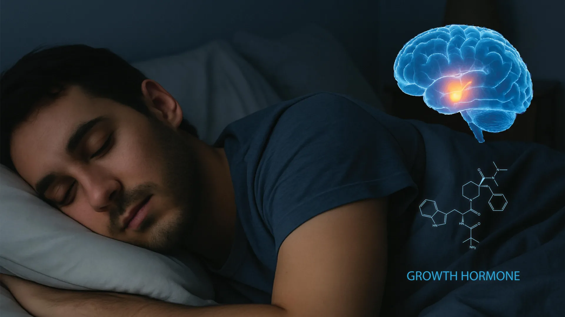
The intricate relationship between the human sleep cycle and the endocrine system has long been observed by the medical community, yet the specific mechanical "switches" within the brain that govern these interactions have remained largely elusive. A landmark study published in the journal Cell by a research team at the University of California, Berkeley, has finally mapped the neural circuitry responsible for the release of growth hormone (GH) during sleep. By identifying the specific neurons and feedback loops involved, the researchers have provided a foundational blueprint that could revolutionize the treatment of metabolic disorders, sleep-related growth deficiencies, and neurodegenerative diseases.
Growth hormone is a critical peptide hormone secreted by the pituitary gland. While it is best known for its role in childhood development and height attainment, its importance persists throughout adulthood. GH is essential for protein synthesis, muscle mass maintenance, bone density, and the regulation of glucose and lipid metabolism. Historically, clinicians have noted that GH levels spike significantly during the onset of deep sleep—specifically during Stage 3 non-rapid eye movement (NREM) sleep—but the "why" and "how" of this neurological orchestration had not been visualized in real-time until now.
The Biological Blueprint of Sleep-Induced Growth
The Berkeley study, led by first author Xinlu Ding and senior author Yang Dan, a professor of neuroscience and molecular and cell biology, focused on the hypothalamus, a region often referred to as the brain’s "command center" for autonomic functions. Within this region, the team isolated the activity of growth hormone-releasing hormone (GHRH) neurons and somatostatin neurons. These two cell types act as a biological "accelerator" and "brake," respectively.
Under normal physiological conditions, GHRH stimulates the pituitary gland to release growth hormone into the bloodstream, while somatostatin acts to inhibit this release. The researchers discovered that the interplay between these neurons is highly sensitive to the specific stages of sleep. Using advanced neural recording techniques in mouse models, the team observed that during REM sleep—the stage associated with vivid dreaming—both GHRH and somatostatin neurons increase their activity. However, during NREM sleep, somatostatin levels drop significantly while GHRH remains moderately active, creating a "permissive window" that allows for the massive pulses of growth hormone characteristic of deep rest.
This discovery is significant because it moves beyond the observational. Previously, scientists relied on serial blood draws to track GH levels, a method that is both invasive and lacks the temporal resolution to match hormone spikes with specific neural firing patterns. By recording neural activity directly, the Berkeley team has provided the first high-resolution map of the brain’s internal endocrine control system.
The Locus Coeruleus and the Discovery of a Novel Feedback Loop
Perhaps the most unexpected finding of the study involves the locus coeruleus (LC), a small nucleus in the brainstem. The LC is the brain’s primary source of norepinephrine, a neurotransmitter associated with wakefulness, attention, and the "fight or flight" response. Conventionally, the LC is thought to be most active during the day and relatively quiet during sleep.
The Berkeley researchers found that when growth hormone is released into the system, it travels back to the brain and activates neurons within the locus coeruleus. This creates a sophisticated feedback loop. As GH levels gradually rise during a period of sleep, the hormone stimulates the LC, which in turn encourages the brain to transition toward wakefulness.
"This suggests that sleep and growth hormone form a tightly balanced system," noted Daniel Silverman, a postdoctoral fellow at UC Berkeley and co-author of the study. "Too little sleep reduces growth hormone release, and too much growth hormone can in turn push the brain toward wakefulness."
Interestingly, the team observed a paradoxical effect: if the activity in the locus coeruleus becomes excessively high, it begins to promote sleepiness rather than alertness. This nuanced self-regulation ensures that the body does not remain in a state of hyper-arousal, maintaining the delicate homeostasis required for both physical repair and cognitive recovery.
Chronology of Research and Methodological Innovations
The road to this discovery involved years of iterative experimentation within Professor Yang Dan’s laboratory. The team utilized optogenetics—a biological technique that involves the use of light to control neurons that have been genetically sensitized to light. By placing micro-electrodes and fiber-optic sensors into the brains of mice, the researchers were able to stimulate specific hypothalamic neurons and observe the immediate hormonal and behavioral consequences.
Mice are ideal subjects for sleep research because, while their sleep-wake cycles are polyphasic (occurring in short bursts throughout a 24-hour period), the underlying neurochemistry and the structure of the hypothalamus are remarkably similar to those of humans. The ability to observe dozens of sleep-wake transitions in a single day allowed the team to gather a robust dataset that would have taken months to collect in human subjects.
The timeline of the research also coincided with a separate study by Silverman earlier this year, which first hinted at the locus coeruleus’s role in promoting sleep under high-stress or high-activity conditions. The convergence of these two lines of inquiry—one focused on hormonal release and the other on arousal circuitry—led to the comprehensive model published in Cell.
Clinical Implications: Metabolism and Cardiovascular Health
The enrichment of our understanding of the GH-sleep circuit has immediate implications for public health, particularly regarding the global rise in metabolic syndrome. Growth hormone is a major regulator of how the body processes fats and sugars. It promotes lipolysis (the breakdown of fats) and helps maintain stable blood glucose levels.
When sleep is chronically fragmented or insufficient—a condition affecting an estimated 30% of the global adult population—the GH-sleep circuit is disrupted. Reduced GH levels lead to decreased fat oxidation and increased insulin resistance. This creates a dangerous cycle where poor sleep contributes to obesity and type 2 diabetes, which in turn makes quality sleep more difficult to achieve due to conditions like sleep apnea.
By identifying the specific GHRH and somatostatin pathways, pharmaceutical researchers may be able to develop targeted therapies that mimic the natural pulsatile release of GH during sleep. This could provide a more effective alternative to current growth hormone replacement therapies, which are often administered via injection and can carry risks of side effects if not perfectly timed with the body’s circadian rhythm.
Neurodegeneration and Cognitive Function
The implications of the Berkeley study extend beyond physical growth and metabolism into the realm of neurology. The locus coeruleus, identified as a key component of the GH feedback loop, is one of the first brain regions to show signs of degeneration in patients with Alzheimer’s and Parkinson’s diseases.
In neurodegenerative conditions, the loss of LC neurons leads to severe sleep disturbances and "sundowning," where patients become agitated or confused in the late afternoon and evening. If growth hormone serves as a modulator for LC excitability, it is possible that maintaining healthy GH levels could help preserve LC function or mitigate some of the cognitive symptoms of these diseases.
Furthermore, the study suggests that GH may play a role in "arousal quality." As Xinlu Ding noted, the hormone may provide cognitive benefits by optimizing alertness levels upon waking. This "refreshing" feeling after a good night’s sleep may be as much a product of hormonal balance as it is of neural rest.
Expert Reactions and Future Directions
The scientific community has responded to the Berkeley study with cautious optimism. Independent researchers note that while the mouse model is highly predictive, human clinical trials will be necessary to confirm the exact thresholds of the GH-LC feedback loop in the human brain.
"The identification of a direct circuit between the hypothalamus and the locus coeruleus via growth hormone is a significant leap forward," said one neuroendocrinologist not involved in the study. "It provides a ‘handle’ for future gene therapies. We are moving away from broad-spectrum drugs and toward cell-specific interventions."
The Berkeley team is already looking toward the next phase of research, which involves investigating how these circuits change with age. As humans age, the amount of deep NREM sleep decreases, as does the production of growth hormone. Determining whether the decline in sleep quality causes the decline in GH, or vice versa, could unlock new strategies for healthy aging and longevity.
The research was supported by prestigious institutions, including the Howard Hughes Medical Institute (HHMI) and the Pivotal Life Sciences Chancellor’s Chair fund. As the medical community continues to digest these findings, the study stands as a testament to the power of circuit-level neuroscience in solving age-old mysteries of human biology. By mapping the invisible threads that connect a quiet night’s sleep to the body’s vital growth processes, the team at UC Berkeley has opened a new door to understanding the holistic nature of human health.
