
 By Lina carolina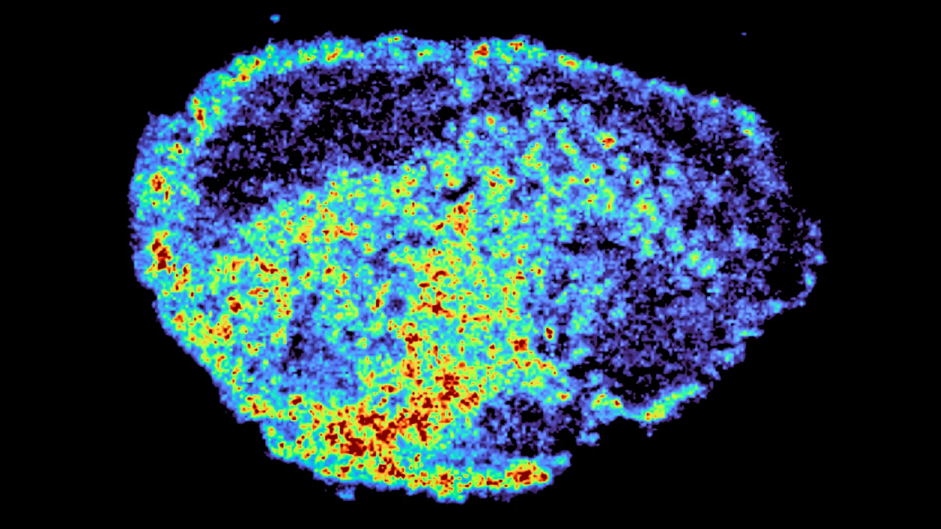
One of the most persistent and frustrating challenges in modern cancer care is the stark variability in treatment response. A therapy that dramatically halts tumor growth in one patient may prove utterly ineffective in another, even when diagnosed with the same cancer type and stage. This fundamental discrepancy has driven intense research efforts to unravel the complex biological mechanisms underlying drug efficacy and resistance. A groundbreaking study, published in the prestigious journal Nature Communications, has shed crucial light on this enigma by focusing on PARP inhibitors, a vital class of targeted therapies, and their intricate journey within ovarian tumor samples. Led by Dr. Louise Fets at the MRC Laboratory of Medical Sciences (LMS), the research team employed cutting-edge imaging techniques to meticulously track the distribution of these drugs, revealing a surprising role for cellular "recycling centers" – lysosomes – in dictating treatment success.
The Mystery of Differential Drug Response
The landscape of cancer treatment has undergone a revolution in recent decades, with targeted therapies offering new hope and significantly improving outcomes for a substantial number of patients. PARP (poly ADP-ribose polymerase) inhibitors, in particular, have been transformative in the management of ovarian cancer, demonstrating remarkable efficacy in patients with BRCA mutations and other DNA repair deficiencies. These drugs work by inhibiting the PARP enzyme, which plays a critical role in DNA repair. In cancer cells with compromised DNA repair pathways (like those with BRCA mutations), blocking PARP leads to an accumulation of DNA damage that ultimately triggers cell death.
However, the clinical reality often falls short of this potential. Not all patients respond to PARP inhibitors, and many who initially benefit develop resistance over time. A core prerequisite for these drugs to exert their cytotoxic effect is their ability to reach and accumulate within cancer cells at sufficiently high concentrations. Despite this fundamental requirement, a comprehensive understanding of how drugs distribute within the complex architecture of tumors and what intracellular mechanisms govern this process has remained elusive. This knowledge gap has hindered the development of strategies to optimize drug delivery and overcome resistance mechanisms.
The research spearheaded by Dr. Fets and her team addresses this critical void. Their findings underscore a paradigm shift in understanding drug efficacy: it is not solely about whether a drug reaches a tumor, but crucially, how it subsequently distributes within the tumor microenvironment and, at a cellular level, within individual cancer cells. To investigate this, the scientists utilized a sophisticated experimental model involving thin slices of human ovarian tumors, meticulously collected from patients and maintained in a viable state ex vivo. These "explants" provided a realistic three-dimensional representation of tumor tissue, allowing researchers to treat them directly with PARP inhibitors and observe the drugs’ movement in real human tumor cells.
Advanced Imaging Unveils Drug Distribution Patterns
The study’s methodological innovation was pivotal. The researchers employed mass spectrometry imaging (MSI), a powerful analytical technique that enables the precise spatial localization and quantification of molecules within biological samples. By applying MSI to the ovarian tumor explants, the team was able to generate highly detailed maps, visually demonstrating the exact locations and concentrations of PARP inhibitors throughout the tumor tissue. This granular mapping provided an unprecedented view of drug uptake and distribution at a level of detail previously unattainable.
Complementing MSI, the researchers utilized spatial transcriptomics. This cutting-edge technology allowed them to simultaneously examine gene expression patterns within the same tissue slice where drug levels were mapped. By comparing gene activity in regions exhibiting high drug accumulation versus those with low drug concentrations, scientists could begin to correlate cellular responses with drug exposure.
The results were striking and revealed significant heterogeneity in drug distribution. Not only did drug levels vary considerably between different patients’ tumors, but even within a single tumor, considerable differences in drug concentration were observed at the cellular and subcellular levels. This variability persisted even when identical doses of the PARP inhibitors were administered to the explants.
"A novel aspect of this study was the use of mass spectrometry imaging to directly measure and visualize drug uptake in patient tumour tissue," stated Dr. Zoe Hall, a senior author of the study and Associate Professor at Imperial’s Department of Metabolism, Digestion and Reproduction. "Through the spatial mapping of drug molecules, we could pinpoint regions of high and low drug and compare gene expression, from the same tissue slice, using spatial transcriptomics." This integrated approach provided a comprehensive picture, linking the physical presence of the drug to the underlying molecular landscape of the tumor.
Lysosomes: The Unseen Drug Reservoirs
The most significant revelation from the study was the central role played by lysosomes in this observed drug distribution heterogeneity. Lysosomes, often described as the "recycling centers" of the cell, are membrane-bound organelles responsible for breaking down waste materials and cellular debris. The research demonstrated that certain PARP inhibitors, upon entering cancer cells, were actively taken up by lysosomes and became sequestered within these organelles. Instead of freely diffusing throughout the cytoplasm, these drugs were essentially trapped, forming internal reservoirs.
This lysosomal sequestration had profound implications for drug availability. The lysosomes acted as slow-release depots, gradually dispensing the accumulated drug back into the cell. This process led to uneven drug exposure: some cells experienced prolonged and higher drug exposure due to their lysosomes’ capacity to store and release the medication, while others, with fewer or less efficient lysosomal storage mechanisms, received significantly lower drug concentrations.
Crucially, the study revealed that this lysosomal trapping was not a universal phenomenon for all PARP inhibitors. The researchers found that drugs such as rucaparib and niraparib were significantly influenced by this mechanism, accumulating within lysosomes. In contrast, other PARP inhibitors, like olaparib, appeared to be less affected by lysosomal sequestration, suggesting distinct intracellular trafficking and accumulation profiles among drugs within the same class.
"We were surprised to see large variability in drug accumulation at the single-cell level," commented Dr. Carmen Ramirez Moncayo, the study’s first author and a Postdoctoral Researcher at the LMS. "This variability was driven by the build-up of a drug in lysosomes, which are acting as reservoirs, increasing the exposure of cancer cells to drugs, by storing and releasing the drug when needed." This observation highlights the complex interplay between drug physicochemical properties and cellular uptake and storage mechanisms.
Implications for Personalized Cancer Therapy
The findings from this study carry substantial weight for the future of cancer treatment, particularly for PARP inhibitors, which are already widely employed in the management of ovarian, breast, and prostate cancers, and are under investigation for numerous other malignancies. A deeper understanding of how these drugs are stored and distributed within cancer cells opens up avenues for developing more precise and personalized therapeutic strategies.
By identifying lysosomal sequestration as a key determinant of drug exposure, clinicians and researchers can begin to tailor treatment approaches to individual patients. This could involve selecting specific PARP inhibitors based on a tumor’s lysosomal characteristics, or potentially developing strategies to modulate lysosomal function to enhance drug delivery and overcome resistance. The ultimate goal is to maximize treatment effectiveness, minimize the development of resistance, and reduce the likelihood of cancer recurrence.
"By understanding how drugs are taken up into cells, we can understand whether this influences why cancer drugs work for some people and not for others," explained Dr. Louise Fets, a senior author and Head of the LMS’ Drug Transport and Tumour Metabolism Group. "Eventually, we hope to be able study the molecular signature of a patient’s tumor to help to tailor therapeutic approaches in a more personalized way." This vision of personalized medicine hinges on precisely mapping the biological factors that govern drug response.
It is important to acknowledge the limitations of the current study. The research was conducted using tumor tissue maintained outside the body. In a living patient, drug delivery occurs via the bloodstream, and the often disorganized and leaky vasculature of tumors can further contribute to uneven drug distribution. Therefore, future research is crucial to bridge the gap between ex vivo findings and the clinical setting.
The next steps for the research team involve employing animal models and expanding studies to larger patient cohorts. These investigations will aim to comprehensively understand the complex interactions between drug delivery mechanisms, tumor architecture, and the influence of lysosomal storage in real-world clinical scenarios, including in the context of relapsed and treatment-resistant cancers. By integrating these findings, the scientific community can move closer to a future where cancer therapies are not only more effective but also precisely tailored to the unique biological profile of each patient’s tumor.
This significant research was made possible through substantial funding from various bodies, including the Medical Research Council, Cancer Research UK, a PhD studentship from the Integrative Toxicology Training Partnership administered by the MRC Toxicology Unit, and a Victoria’s Secret Global Fund for Women’s Cancers Career Development Award, in partnership with Pelotonia and AACR. These collaborations underscore the multi-faceted support required for advancing complex cancer research.
