
 By Nana O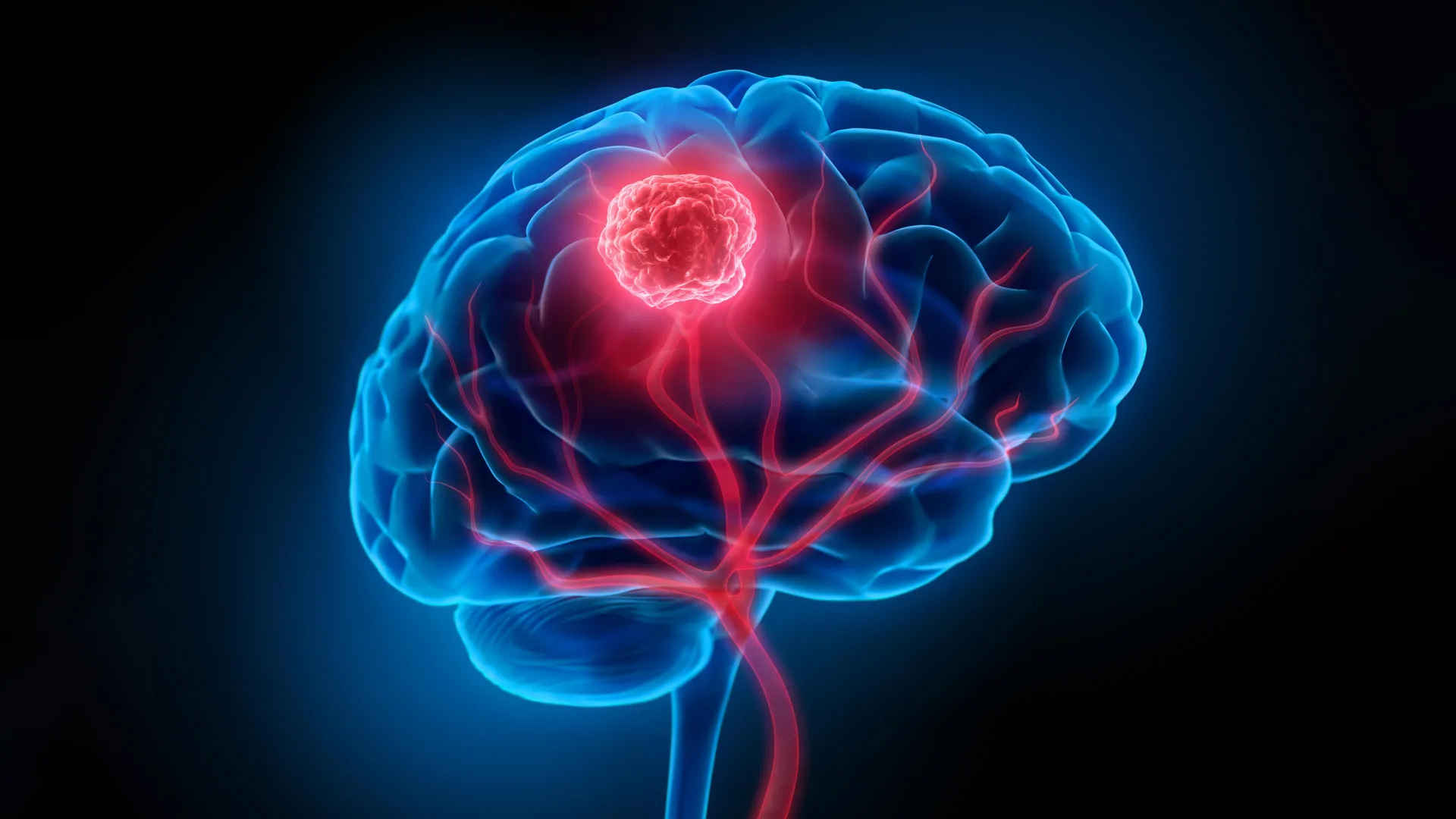
A groundbreaking study from South Korea is poised to redefine our understanding of IDH-mutant glioma, the most prevalent and aggressive malignant brain tumor affecting adults under 50. For decades, the prevailing treatment strategy has revolved around surgically removing the visible tumor mass. However, new research published in the prestigious journal Science on January 8th suggests this approach may be overlooking the earliest, most insidious stages of the disease, potentially explaining the high rates of recurrence that plague patients. Scientists have discovered that normal-looking brain cells can acquire the initial IDH mutation and stealthily disseminate throughout the brain’s intricate cortex, long before any detectable tumor mass materializes. This hidden, nascent phase offers a compelling explanation for the cancer’s tenacious resistance to therapy and illuminates novel avenues for earlier detection and the prevention of relapse.
Tracing the Genesis: From Normal Cells to Malignant Growth
The pivotal findings emerged from a collaborative research effort spearheaded by Professor Jeong Ho Lee of the KAIST Graduate School of Medical Science and Engineering and Professor Seok-Gu Kang of Yonsei University Severance Hospital. Their investigation pinpointed the cellular origin of IDH-mutant glioma, revealing that these devastating tumors originate not from abnormal cells but from Glial Progenitor Cells (GPCs) that are integral components of healthy brain tissue.
Glial Progenitor Cells (GPCs) are a critical element of the brain’s cellular landscape. These versatile cells possess the remarkable ability to differentiate into various types of glial cells, which are essential for supporting and protecting neurons. In the context of IDH-mutant glioma, these GPCs, when subjected to specific genetic alterations, can initiate a cascade of events leading to uncontrolled proliferation and tumor formation.
To substantiate their hypothesis, the research team undertook an exhaustive examination of tumor samples procured during extensive surgical interventions. Crucially, they also analyzed adjacent brain tissue that, to the naked eye, appeared entirely healthy. Their meticulous scrutiny unveiled a startling reality: cells harboring the IDH mutation were demonstrably present within brain regions that showed no outward signs of abnormality. This observation provided the first definitive evidence that the genesis of malignant brain tumors is not a sudden event but rather a protracted process that commences discreetly within seemingly normal brain tissue, evolving gradually over many years before coalescing into a clinically detectable mass.
Technological Prowess Illuminates Early Cellular Events
Confirming the identity of these early mutated cells necessitated the application of cutting-edge scientific methodologies. The researchers employed "spatial transcriptomics," an advanced analytical technique that allows for the simultaneous mapping of gene expression patterns within specific spatial locations in tissue. This powerful tool enabled them to precisely determine "which genes are operating where" within the brain samples. The results unequivocally confirmed that the mutation-bearing cells were indeed Glial Progenitor Cells (GPCs) located within the cerebral cortex, the outermost layer of the brain responsible for higher-level cognitive functions.
To further validate their findings and replicate the developmental trajectory of these tumors, the team engineered an animal model. By introducing the same critical genetic "driver mutation" identified in human patients into the GPCs of mice, they successfully recapitulated key stages of brain tumor development. This experimental approach provided invaluable insights into the step-by-step process by which normal GPCs transform into cancerous cells.
A Dichotomy in Brain Cancer Development: New Insights into Subtype Specificity
This latest research builds upon a significant prior discovery made by Professor Lee’s group. In 2018, they reported that IDH wildtype glioblastoma, another highly aggressive and often fatal brain cancer, originates from neural stem cells residing in the subventricular zone, a region known for generating new brain cells in adults. This earlier work, published in the esteemed journal Nature, established a precedent for understanding the cellular origins of different brain cancer subtypes.
The current study’s findings highlight a crucial distinction: while both IDH wildtype glioblastoma and IDH-mutant glioma are devastating malignant brain tumors, they arise from distinct cell types and initiate in different anatomical locations within the brain. This comparative analysis reinforces the understanding that brain cancers are not monolithic entities but rather follow diverse biological pathways, influenced by their specific genetic mutations and cellular origins. This realization is paramount for developing targeted and effective therapeutic interventions.
Transforming the Landscape of Early Diagnosis and Recurrence Suppression
The implications of this paradigm-shifting research are profound, particularly for the fields of early cancer detection and the prevention of tumor recurrence. Professor Seok-Gu Kang, a co-corresponding author on the study, emphasized the significance of this new understanding. "Brain tumors may not start exactly where the tumor mass is visible," he stated. "A targeted approach focused on the origin cells and the site of origin according to the brain tumor subtype will serve as a crucial clue to changing the paradigm of early diagnosis and recurrence suppression treatment."
This insight suggests that current diagnostic methods, which primarily rely on identifying visible tumor masses through imaging techniques like MRI or CT scans, may be too late in detecting the disease. By identifying the earliest cellular precursors of IDH-mutant glioma, researchers and clinicians can now explore novel strategies for detecting the disease at its nascent stages, potentially before it becomes clinically apparent and more challenging to treat.
Furthermore, understanding the initial mutational events and the spread of these mutated cells within the brain offers new hope for preventing recurrence. The persistent presence of microscopic disease, even after seemingly successful tumor removal, is a major contributor to relapse. By targeting these early-stage, disseminated cells, therapeutic strategies could be developed to eradicate the cancer more comprehensively.
From Discovery to Therapeutic Innovation: A Multi-pronged Approach
In direct response to these groundbreaking findings, tangible steps are already underway to translate this scientific knowledge into clinical practice. Sovagen Co., Ltd., a faculty startup originating from KAIST, is actively developing an innovative RNA-based drug. This novel therapeutic agent is designed to specifically target and inhibit the progression and recurrence of IDH-mutant malignant brain tumors. The development of such targeted therapies represents a significant departure from traditional, broadly cytotoxic chemotherapy regimens.
Concurrently, Severance Hospital is spearheading the development of advanced technologies aimed at detecting and controlling these early-stage mutant cells. This initiative is being pursued through the Korea-US Innovative Result Creation R&D project, fostering international collaboration and accelerating the translation of research into tangible clinical solutions. The focus on early detection and control underscores a proactive approach to combating this aggressive disease.
The Spark of Inquiry: A Surgeon’s Persistence Fuels Discovery
The journey from fundamental scientific inquiry to clinical application was fueled by a deep-seated question that resonated with one of the study’s lead investigators. Dr. Jung Won Park, a neurosurgeon and the study’s sole first author, who is also a postdoctoral researcher at KAIST Graduate School of Medical Science and Engineering, highlighted the synergistic collaboration that underpinned this achievement. "This achievement was made possible by combining KAIST’s world-class basic science research capabilities with the clinical expertise of Yonsei Severance Hospital," Dr. Park remarked. He further elaborated on the genesis of the research, stating, "The question I kept asking while treating patients – ‘Where does this tumor originate?’ – was the starting point of this research." This sentiment underscores the critical role of clinician-scientists in bridging the gap between laboratory discoveries and real-world patient care, driven by the direct experiences of confronting challenging diseases.
A Glimpse into the Broader Scientific Landscape
The successful publication of these findings in Science signifies not only a triumph for the research teams involved but also a substantial advancement for the broader scientific community engaged in neuro-oncology research. The study received crucial financial support from a consortium of esteemed organizations, including the Suh Kyung-bae Science Foundation, the National Research Foundation of Korea, the Ministry of Science and ICT, the Ministry of Health and Welfare, and the Korea Health Industry Development Institute (through its Physician-Scientist Training Program). This multifaceted support highlights the national and international recognition of the importance and potential impact of this research.
The implications of this research extend beyond IDH-mutant glioma. The methodological advancements, particularly in spatial transcriptomics and animal modeling, can be readily applied to the study of other complex diseases. The identification of distinct cellular origins for different brain tumor subtypes also paves the way for more personalized medicine approaches, where treatments can be tailored to the specific biological characteristics of a patient’s tumor.
The discovery that normal brain cells can harbor the initial genetic mutations for cancer, and that these mutations can spread long before a visible tumor forms, presents a significant challenge and opportunity. It necessitates a re-evaluation of diagnostic paradigms, moving beyond the detection of gross pathology to the identification of subtle molecular signatures at the cellular level. It also opens up new frontiers for preventative strategies, potentially intervening at the earliest stages of cellular transformation to halt disease progression before it takes hold.
As the scientific community digests these findings, the focus will likely shift towards developing and validating new diagnostic tools, refining targeted therapies, and exploring the feasibility of early intervention strategies. The path forward, illuminated by this pioneering research, promises a more nuanced and effective approach to confronting the formidable challenge of IDH-mutant glioma and, potentially, other complex cancers. The initial spark of a surgeon’s persistent question has ignited a wave of discovery that could fundamentally alter the prognosis for countless patients worldwide.
