
 By Lina carolina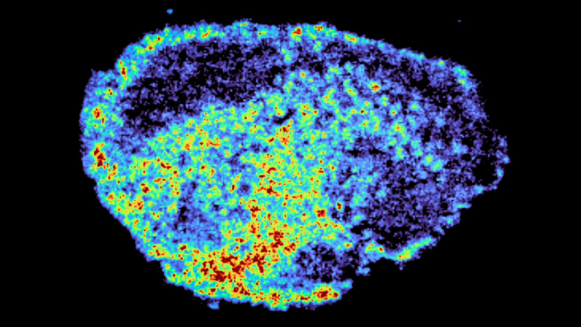
One of the most persistent and vexing challenges in the field of oncology is the stark variability in patient response to cancer therapies. A single treatment, heralded for its promise, can be a life-saving intervention for some individuals, while proving utterly ineffective for others. This perplexing disparity, particularly evident with targeted therapies, has long been a focus of intense scientific scrutiny. Now, groundbreaking research published in the esteemed journal Nature Communications offers a significant leap forward in understanding this phenomenon, shedding light on the intricate cellular mechanisms that dictate drug efficacy.
Led by Dr. Louise Fets, a prominent researcher at the Medical Research Council (MRC) Laboratory of Medical Sciences (LMS), the study meticulously investigates the behavior of PARP inhibitors, a crucial class of targeted drugs widely employed in cancer treatment. Utilizing sophisticated advanced imaging techniques, the research team has for the first time precisely tracked the journey of these powerful molecules within ovarian tumor samples, revealing a previously underestimated role for cellular organelles known as lysosomes.
The Cellular Nexus: Lysosomes as Unseen Drug Compartments
The core finding of this pivotal study is that PARP inhibitors, far from distributing uniformly, can accumulate within lysosomes. These organelles, often described as the cell’s "recycling centers," play a vital role in breaking down cellular waste products and engulfed materials. However, in the context of cancer therapy, the research demonstrates that lysosomes can act as unintended drug reservoirs. Once inside these compartments, the drugs can become sequestered, influencing their availability and subsequent release, thereby profoundly impacting the overall effectiveness of the treatment.
This discovery directly addresses a critical knowledge gap: while the importance of a drug reaching its target and achieving sufficient concentration within cancer cells to induce cell death is understood, the precise dynamics of drug distribution within tumors and individual cells have remained largely elusive. The implications are substantial, suggesting that treatment success hinges not solely on the drug’s initial arrival at the tumor site, but also on its complex intracellular trafficking and localization.
Mapping the Intracellular Landscape: Advanced Imaging Reveals Drug Dynamics
In recent years, the landscape of cancer treatment has been revolutionized by the rapid development of novel therapeutic agents, offering renewed hope and improved outcomes for countless patients. PARP inhibitors, in particular, have marked a transformative era in the management of ovarian cancer, demonstrating remarkable efficacy in many cases. Nevertheless, the persistent challenge of non-response and the emergence of drug resistance over time underscore the need for deeper comprehension of their mechanisms.
To address this, Dr. Fets and her team employed a novel approach, utilizing patient-derived ovarian tumor samples. These meticulously preserved "explants" – living tumor tissue maintained ex vivo – were treated with PARP inhibitors. This innovative methodology allowed researchers to observe the drugs’ behavior in a realistic human tumor microenvironment, providing an unprecedented level of detail.
The study’s technological prowess was central to its success. The researchers integrated mass spectrometry imaging with spatial transcriptomics. Mass spectrometry imaging provided high-resolution maps, precisely delineating the locations where PARP inhibitors accumulated within the tumor tissue. This spatial information was then correlated with spatial transcriptomics data, which allowed for the examination of gene expression patterns in areas of both high and low drug concentration within the same tissue slice. The resulting comprehensive analysis unveiled striking heterogeneity in drug distribution, not only between different tumors but also between individual patients, even when subjected to identical dosages.
Dr. Zoe Hall, a senior author of the study and Associate Professor at Imperial College London’s Department of Metabolism, Digestion and Reproduction, highlighted the innovative nature of their methodology. "A novel aspect of this study was the use of mass spectrometry imaging to directly measure and visualize drug uptake in patient tumour tissue," Dr. Hall stated. "Through the spatial mapping of drug molecules, we could pinpoint regions of high and low drug and compare gene expression, from the same tissue slice, using spatial transcriptomics." This dual approach provided a holistic view, linking drug localization with cellular responses at a granular level.
Lysosomal Sequestration: A Hidden Mechanism of Drug Variability
The findings unequivocally point to lysosomes as central players in the observed uneven drug distribution. The study observed that certain PARP inhibitors exhibit a propensity to be internalized by lysosomes, where they are then sequestered rather than diffusing freely throughout the cell. This phenomenon effectively creates intracellular "pockets" of concentrated drug.
These lysosomal accumulations function as slow-release reservoirs. By retaining and gradually releasing the drug, lysosomes can lead to prolonged exposure in some cells, while others, situated in different regions of the tumor or possessing different lysosomal characteristics, receive significantly lower drug concentrations. This differential exposure can have a profound impact on therapeutic outcomes.
Crucially, the study revealed that this lysosomal sequestration is not a universal behavior for all PARP inhibitors. While drugs such as rucaparib and niraparib were found to be significantly influenced by this lysosomal trapping mechanism, another widely used PARP inhibitor, olaparib, appeared to be largely unaffected. This distinction suggests that the chemical properties of individual drugs play a critical role in their interaction with cellular organelles like lysosomes.
Dr. Carmen Ramirez Moncayo, the study’s first author and a Postdoctoral Researcher at the LMS, expressed her surprise at the extent of this intracellular variability. "We were surprised to see large variability in drug accumulation at the single-cell level," Dr. Ramirez Moncayo commented. "This variability was driven by the build-up of a drug in lysosomes, which are acting as reservoirs, increasing the exposure of cancer cells to drugs, by storing and releasing the drug when needed." This observation underscores the dynamic and complex nature of drug-cell interactions, extending beyond simple uptake and diffusion models.
Charting a Course for Personalized Cancer Therapy
The clinical significance of these findings cannot be overstated. PARP inhibitors are currently a cornerstone of treatment for ovarian, breast, and prostate cancers, and their therapeutic potential is being actively explored in a growing number of other cancer types. A more profound understanding of how these drugs are stored and distributed within the cellular milieu opens up exciting avenues for developing more personalized and effective treatment strategies. By accounting for individual variations in drug pharmacokinetics and pharmacodynamics, clinicians may be able to optimize drug selection, dosage, and timing, thereby enhancing treatment efficacy, mitigating the development of resistance, and ultimately reducing the rates of cancer relapse.
Dr. Louise Fets, a senior author and Head of the LMS’ Drug Transport and Tumour Metabolism Group, articulated the long-term vision stemming from this research. "By understanding how drugs are taken up into cells, we can understand whether this influences why cancer drugs work for some people and not for others," Dr. Fets explained. "Eventually, we hope to be able study the molecular signature of a patient’s tumor to help to tailor therapeutic approaches in a more personalized way." This aspiration points towards a future where cancer treatment is not a one-size-fits-all approach, but a precisely tailored intervention informed by a deep understanding of individual tumor biology.
It is important to acknowledge the context of the study’s methodology. The research was conducted using tumor tissue maintained outside the body. In a living patient, drug delivery is facilitated by the bloodstream, and tumor vasculature is often notoriously disorganized and aberrant. These factors can further complicate drug distribution, potentially exacerbating the unevenness observed in the explant models. Future research will therefore be crucial in bridging this gap. Studies employing animal models and larger patient cohorts will be essential to elucidate how drug delivery mechanisms, the intricate architecture of tumor blood vessels, and the dynamics of lysosomal drug storage interact within the complex clinical setting, particularly in the context of relapsed or refractory cancers. Understanding these in-vivo dynamics will be paramount to translating these groundbreaking laboratory findings into tangible clinical benefits.
This research was made possible through substantial support from various funding bodies, including the Medical Research Council, Cancer Research UK, a PhD studentship from the Integrative Toxicology Training Partnership administered by the MRC Toxicology Unit, and a Victoria’s Secret Global Fund for Women’s Cancers Career Development Award, in partnership with Pelotonia and the American Association for Cancer Research (AACR). The collaborative nature of this funding underscores the widespread recognition of the importance of this research in advancing cancer care.
