
 By Suherman Candra Maholtra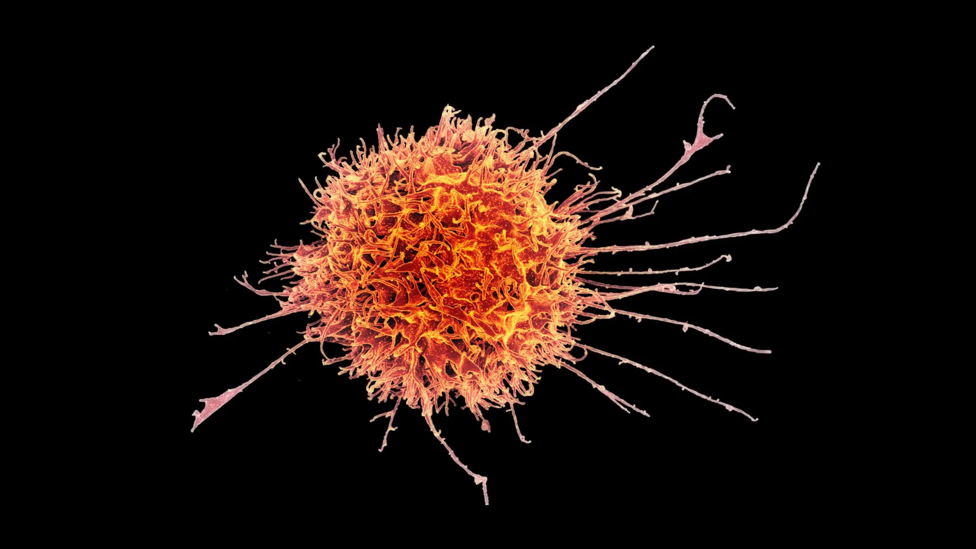
While cell-based immunotherapies have achieved landmark successes in treating hematologic malignancies, such as leukemia and lymphoma, the therapeutic landscape for solid tumors has remained stubbornly resistant to similar breakthroughs. Solid tumors, which account for the vast majority of adult cancer deaths, present a formidable fortress of physical and chemical barriers. These masses are often impenetrable to circulating immune cells and frequently emit immunosuppressive signals that disarm any white blood cells that manage to enter. However, a new study led by researchers at Stanford Medicine and published in Science Translational Medicine has unveiled a sophisticated strategy to breach these defenses. By reprogramming natural killer (NK) cells into a specialized "tissue-resident" state, the team has developed a method to enable these cells to infiltrate solid tumors and execute targeted destruction of malignant tissue.
The research, led by John Sunwoo, MD, the Edward C. and Amy H. Sewall Professor in the School of Medicine, marks a significant pivot in the field of oncology. Historically, immunotherapy has relied heavily on T cells, which require complex genetic engineering and are often patient-specific. The Stanford approach focuses on the innate immune system’s "first responders"—natural killer cells—and utilizes a biological "recipe" to transform them into highly aggressive, tumor-infiltrating agents.
The Evolution of Natural Killer Cell Research
Natural killer cells were first identified in the 1970s, distinguished by their innate ability to recognize and destroy virally infected or cancerous cells without prior sensitization. Unlike T cells, which must be "primed" to recognize specific antigens, NK cells monitor the body for general signs of cellular stress or the absence of "self" markers. Despite their potency in the bloodstream, their efficacy against solid tumors has been historically limited by their inability to take up permanent residence within the dense, hostile environment of a tumor.
Most immunological research over the last half-century has focused on "circulating" immune cells—those that travel through the blood and lymph. However, the Stanford team recognized that the most effective immune responses often occur within the tissues themselves. Tissue-resident NK (trNK) cells are naturally found in the skin, liver, and lungs, where they act as localized sentinels. The challenge for the researchers was to understand why some trNK cells act as aggressive killers while others appear to be immunosuppressive or "exhausted."
"For a long time, the study of immunology and disease in humans was concentrated on the blood immune cells," Sunwoo explained. "With the advancement of tools and bioinformatics, we are now starting to look more at what’s going on in tissue. For most immune cells, the tissue is where the action is."
Cracking the "Goldilocks" Code of TGF-beta
The breakthrough hinged on deciphering the role of Transforming Growth Factor beta (TGF-b), a signaling protein ubiquitous in the tumor microenvironment. TGF-b is a double-edged sword in biology; it is essential for cell development but is also frequently hijacked by tumors to suppress immune activity.
Through a series of controlled experiments, Sunwoo’s team isolated circulating NK cells from healthy human donors and exposed them to various concentrations of TGF-b. They discovered a "Goldilocks" effect: the timing and dosage of the signal determined the cell’s ultimate fate. If the NK cells were exposed to too much TGF-b for a prolonged period, they became dysfunctional and suppressed the immune response—a state often seen in the uterine lining during pregnancy to protect the fetus.
Conversely, the researchers found that a brief, high-intensity burst of TGF-b, delivered via direct physical contact with epithelial tumor cells, acted as a maturation switch. This specific interaction transformed the circulating NK cells into a potent, tissue-resident form. These modified cells expressed a specific molecular signature—including surface proteins CD49a, CD103, and crucially, CD39—that allowed them to bind to tumor tissues and maintain their "killer" status.
Molecular Mechanisms of Enhanced Cytotoxicity
The engineered tissue-resident NK cells were found to be significantly more lethal than their circulating counterparts. Detailed molecular analysis revealed that these cells were packed with an arsenal of cytotoxic proteins. Specifically, the researchers observed elevated levels of perforin, a protein that acts like a molecular drill to create pores in the membranes of cancer cells. Once the pores are established, the NK cells deliver granzyme A, a toxic enzyme that triggers programmed cell death (apoptosis) from within the tumor cell.
This high concentration of "molecular machinery" ensures that once a trNK cell infiltrates a tumor, it can neutralize multiple targets in rapid succession. The presence of CD39 was identified as a key marker for this aggressive phenotype, distinguishing the "super-killers" from the suppressed versions of tissue-resident cells.
Preclinical Results: Synergistic Effects with Monoclonal Antibodies
To validate their findings, the Stanford team tested the modified cells in mouse models of human melanoma and head and neck squamous cell carcinoma (HNSCC). The results were highly reproducible: the tissue-resident NK cells successfully migrated into the solid tumors and slowed their growth significantly more than conventional NK cells.
The study also explored the potential for combination therapy. When the engineered NK cells were paired with cetuximab—a monoclonal antibody that targets the Epidermal Growth Factor Receptor (EGFR)—the results were even more pronounced. Cetuximab acts as a beacon, marking cancer cells for destruction, while the trNK cells provide the "muscle" to carry out the attack.
In the mouse trials, a single combined dose of trNK cells and cetuximab led to a dramatic reduction in tumor volume. By day 30 of the study, the mice receiving the combination therapy remained healthy, while the control groups showed significant disease progression. "It was very reproducible, very striking and very clear," Sunwoo noted, emphasizing that the combination therapy overcame the limitations of using cetuximab as a monotherapy, which often yields modest results in human patients.
The "Off-the-Shelf" Advantage: Economic and Clinical Implications
One of the most transformative aspects of this research is its potential to democratize cell therapy. Currently, the most prominent cell therapies, such as CAR-T, are "autologous," meaning they must be manufactured using a patient’s own cells. This process is prohibitively expensive, often costing hundreds of thousands of dollars per patient, and involves a weeks-long manufacturing delay that some advanced-stage cancer patients cannot afford to wait through.
In contrast, natural killer cells are "allogeneic," meaning they do not typically cause graft-versus-host disease when transferred between individuals. This allows for a "universal donor" model. The Stanford team has developed and applied to patent a method to scale the production of these cells. According to their data, a single blood donation could yield enough material to produce approximately 20 doses of modified trNK cells within two weeks.
"It would be almost an off-the-shelf drug," Sunwoo said. "They’ll be cryopreserved, so we can make a bunch of doses and give it to different patients. There would be no delay." This scalability could drastically reduce the cost of treatment and make advanced immunotherapy accessible to community hospitals rather than just elite academic medical centers.
Chronology of Development and Future Directions
The journey from the initial identification of NK cells in the 1970s to this recent breakthrough reflects a steady progression in biotechnological capabilities:
- 1970s: Discovery of NK cells and their role in the innate immune system.
- 2000s-2010s: Identification of TGF-b as a major regulator of the tumor microenvironment.
- 2018-2022: Stanford researchers begin isolating the specific signaling requirements for tissue residency in immune cells.
- 2023: Publication of the "Goldilocks" TGF-b findings in Science Translational Medicine.
- Late 2024 (Projected): Commencement of Phase I clinical trials in humans.
The research team, which includes co-lead authors Nina Horowitz, PhD, Imran Mohammad, PhD, and June Ho Shin, PhD, is now focused on transitioning these findings into the clinic. They are currently seeking Food and Drug Administration (FDA) approval to begin a Phase I trial targeting patients with advanced squamous cell carcinoma. This trial will evaluate the safety and preliminary efficacy of the trNK-cetuximab combination in humans.
Broader Impact on the Oncology Landscape
The implications of this study extend beyond head and neck cancer. The ability to "program" immune cells to prefer tissue environments over the bloodstream could be applied to a wide range of solid tumors, including those of the breast, colon, and pancreas. Furthermore, the discovery that physical contact between cells is necessary for optimal activation suggests new avenues for "biomimetic" manufacturing, where synthetic surfaces are used to simulate the tumor environment during the cell-culturing process.
Independent experts in the field suggest that if the Phase I trials prove successful, this could represent a "third wave" of immunotherapy—moving from non-specific checkpoints to engineered T cells, and finally to highly adaptable, off-the-shelf innate immune cells.
While Sunwoo cautions that mouse models are only a "proof of concept" and that human biology presents unique challenges, the clarity of the data offers a rare sense of optimism in the fight against solid tumors. By turning a tumor’s own signaling molecules against it, the Stanford team may have found the key to unlocking the body’s natural defenses in the places they are needed most.
The study was supported by the National Institutes of Health, the Tai Tsun Wu Research Fund for Natural Killer Cell Immunotherapy, and the Stanford Bio-X Fellowship, with contributions from researchers at Ohio State University and Washington University School of Medicine.
