
 By Billy Candra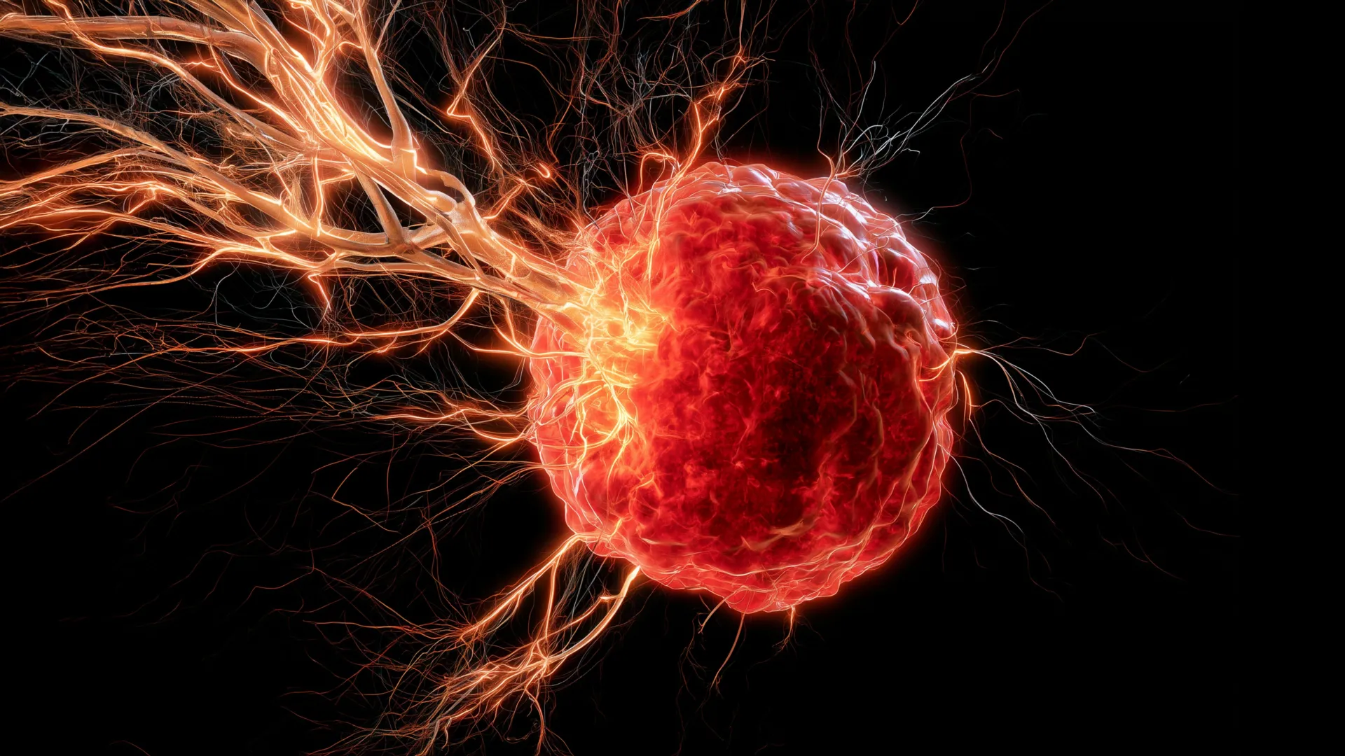
New research emanating from the University of Oklahoma has illuminated a previously unclear mechanism by which an aggressive form of breast cancer, specifically triple-negative breast cancer (TNBC), adeptly manipulates the body’s immune system. This manipulation leads to the recruitment and growth of nerves directly into tumors, a process that appears to foster cancer progression and resistance to therapeutic interventions. This groundbreaking study, recently published in the esteemed journal Cell Death & Differentiation, offers a crucial explanation for how these extensive nerve networks become integrated within solid tumors, a phenomenon long observed but poorly understood.
The Enigma of Tumor Innervation: A Long-Standing Question
For decades, scientists have recognized the presence of intricate nerve networks within many solid tumors. While the existence of these nerves has been a known histological feature, the precise mechanisms governing their entry and proliferation within the hostile tumor microenvironment have largely remained an enigma. The implications of tumor innervation are profound, with emerging evidence suggesting that these nerve fibers are not merely passive bystanders but active participants in various aspects of cancer biology, including growth, metastasis, and even pain perception associated with the disease. Understanding how these nerves are recruited is therefore critical to unraveling key aspects of cancer pathogenesis.
Triple-negative breast cancer, the focus of the University of Oklahoma study, represents a particularly challenging subtype of breast cancer. Characterized by the absence of estrogen receptor (ER), progesterone receptor (PR), and human epidermal growth factor receptor 2 (HER2) expression, TNBC lacks the specific molecular targets that have revolutionized treatment for other breast cancer types. Consequently, patients with TNBC often face a more aggressive disease course, higher rates of recurrence, and a poorer prognosis, primarily relying on chemotherapy, surgery, and radiation therapy. The average 5-year survival rate for localized TNBC is around 90%, but this drops significantly to approximately 12% for metastatic disease, underscoring the urgent need for novel therapeutic strategies. It is against this backdrop of clinical urgency that the findings from Dr. Maureen Cox’s team gain significant relevance.
Macrophages: A Double-Edged Sword in the Tumor Microenvironment
The research pinpoints macrophages, a type of immune cell renowned for its role in fighting infections and repairing damaged tissues, as central orchestrators in this nerve recruitment process. In healthy physiological contexts, macrophages are vital components of innate immunity, acting as cellular "scavengers" that engulf pathogens and cellular debris, while also mediating inflammatory responses and tissue remodeling. However, within the complex and often perverse ecosystem of a tumor, these normally beneficial immune cells can be hijacked and reprogrammed to serve the cancer’s agenda.
The study reveals that once macrophages infiltrate the tumor microenvironment, they undergo a phenotypic shift, transitioning from their anti-tumorigenic roles to become pro-tumorigenic actors. In this subverted state, they begin to secrete brain-derived neurotrophic factor (BDNF). Dr. Maureen Cox, Ph.D., an assistant professor in the Department of Microbiology and Immunology at the OU College of Medicine and a research member of OU Health Stephenson Cancer Center, elaborated on this critical finding: "Macrophages are the critical source for drawing nerves into the tumor. Although macrophages typically play a positive role in the body, they are facilitating a negative function in this scenario of breast cancer." This statement highlights the insidious nature of cancer’s ability to co-opt the body’s protective mechanisms for its own nefarious purposes. The discovery of macrophages acting as "master manipulators" in nerve recruitment represents a significant advance in understanding the intricate cellular cross-talk within the tumor microenvironment.
Brain-Derived Neurotrophic Factor (BDNF): A Misguided Signal
Brain-derived neurotrophic factor (BDNF) is a well-established neurotrophin, a family of proteins that support the survival, development, and function of neurons. In the central and peripheral nervous systems, BDNF is crucial for neurogenesis, neuronal differentiation, synaptic plasticity, and overall neuronal health. Its role in learning, memory, and mood regulation is extensively documented. However, the University of Oklahoma study has uncovered a sinister twist to this vital biological signal.
The research demonstrates that breast cancer tumors exploit this fundamental neurotrophic pathway. By inducing macrophages to release BDNF, the tumors essentially send out a potent "grow here" signal to nearby nerves, compelling them to extend their processes toward and into the cancerous mass. This hijacking of a conserved neurotrophic pathway by cancer cells underscores the evolutionary adaptability and cunning of malignant processes. The presence of these cancer-associated nerves (CANs) is increasingly linked to aggressive tumor behavior. For instance, studies have shown that nerves can provide direct communication channels for cancer cells, potentially facilitating local invasion and distant metastasis. They can also secrete their own neurotrophic factors and neurotransmitters, creating a feedback loop that further stimulates tumor growth and survival, and potentially even contributing to resistance to conventional therapies.
The Research Journey: From Hypothesis to Validation
The path to this discovery involved a meticulous and systematic research approach led by Dr. Cox and her colleagues. The initial hypothesis posited that there must be specific molecular signals guiding nerve infiltration into tumors, moving beyond the mere observation of their presence.
The team embarked on a series of rigorous experiments, beginning with in vitro studies to identify potential cellular and molecular culprits. These investigations quickly pointed toward the critical involvement of macrophages and their secretory products. Further validation was sought in sophisticated murine (mouse) models of triple-negative breast cancer. In these models, researchers could precisely control experimental conditions and observe the dynamic interplay between immune cells, nerves, and tumor cells in a living system.
A pivotal moment in the research was the experimental intervention where the team utilized a drug designed to block BDNF signaling. The results were striking and highly encouraging: in the mice treated with the BDNF blocking agent, the growth of nerves into the tumors was effectively halted. More importantly, this blockade of nerve infiltration correlated with a significant reduction in overall tumor growth. This direct causal link between BDNF-mediated nerve recruitment and tumor progression provides compelling evidence for the therapeutic potential of targeting this pathway.
Dr. Cox highlighted the immediate translational promise of this finding: "It looks really promising that we can use this drug, which is already on the market, to target BDNF." The potential to repurpose an existing pharmaceutical agent, which has already undergone extensive safety testing and regulatory approval for other indications, significantly accelerates the timeline for potential clinical application. This "fast-track" potential is a major advantage in oncology, where the development of new drugs is a lengthy and costly process. Furthermore, Dr. Cox speculated on another critical implication: "We believe that the nerves are immunosuppressive, so if we can stop the nerves from growing in the first place, maybe we can boost the immune response to help fight the cancer." This suggests a synergistic therapeutic strategy where blocking nerve infiltration could not only directly impede tumor growth but also enhance the efficacy of immunotherapies, which have shown promise but variable success in TNBC.
Translational Insights: Evidence from Human Patients
To bridge the gap between preclinical findings in mouse models and clinical relevance in human disease, the researchers meticulously examined retrospective data from patients diagnosed with triple-negative breast cancer. This crucial step involved analyzing tumor biopsies and clinical outcomes to determine if the biological pattern observed in mice held true for humans.
The analysis revealed a compelling correlation: patients whose tumors exhibited higher levels of both macrophages and BDNF were statistically linked with poorer survival outcomes. This direct association provides robust evidence that the macrophage-BDNF-nerve axis is not merely an experimental artifact in animal models but a clinically significant mechanism operating within human TNBC. This translational validation is paramount for any scientific discovery aiming to impact patient care. It strengthens the argument for pursuing this pathway as a viable therapeutic target and offers a potential prognostic biomarker – elevated levels of macrophages and BDNF could indicate a more aggressive disease course.
The Landscape of Triple-Negative Breast Cancer: A Persistent Challenge
Triple-negative breast cancer accounts for approximately 10-15% of all breast cancer diagnoses, translating to tens of thousands of new cases annually in the United States alone. While less common than other subtypes, its aggressive nature, propensity for early metastasis, and higher recurrence rates make it disproportionately responsible for breast cancer mortality. Unlike hormone receptor-positive or HER2-positive breast cancers, which benefit from targeted endocrine therapies or anti-HER2 drugs, TNBC lacks these specific receptors, rendering these highly effective treatments obsolete.
Current standard-of-care for TNBC primarily involves surgery, chemotherapy (often dose-dense or neoadjuvant), and radiation therapy. While these treatments can be effective, particularly in early-stage disease, they are often associated with significant side effects and are less successful in metastatic settings. The advent of immunotherapy, particularly PD-L1 inhibitors, has offered a new ray of hope for a subset of TNBC patients, but response rates remain suboptimal, and predictive biomarkers for immunotherapy response are still being refined. The University of Oklahoma’s discovery, by identifying a novel vulnerability in TNBC, holds the potential to expand the therapeutic arsenal, either as a standalone treatment or, more likely, in combination with existing or emerging therapies.
Broader Implications and a Paradigm Shift in Oncology
The findings from Dr. Cox’s team represent more than just an incremental advance; they point towards a significant paradigm shift in how cancer is conceptualized and treated. For decades, cancer therapy primarily focused on directly killing cancer cells through cytotoxic agents or radiation. While effective, this approach often comes with collateral damage to healthy cells and can lead to resistance mechanisms. The emerging understanding of the tumor microenvironment (TME) – the complex ecosystem of cells, extracellular matrix, and signaling molecules surrounding a tumor – has revealed that cancer is not just a disease of rogue cells, but a systemic illness deeply intertwined with its surrounding environment.
Targeting components of the TME, such as immune cells or nerve infiltration, offers a sophisticated alternative to direct cell killing. This research suggests that disrupting the communication lines within the TME, specifically the macrophage-BDNF-nerve axis, could be a highly effective strategy. This approach aligns with the growing field of "neuro-oncology," which investigates the intricate interplay between the nervous system and cancer progression. Evidence is accumulating that nerves can influence angiogenesis (blood vessel formation, crucial for tumor nutrient supply), modulate immune responses within the tumor, and even serve as conduits for metastatic spread.
The potential to repurpose an existing drug that targets BDNF signaling is particularly exciting. This could dramatically shorten the time and reduce the cost associated with bringing a new therapy to patients. Moreover, combining a BDNF blocker with current immunotherapies could create a synergistic effect, potentially overcoming resistance mechanisms and boosting the overall anti-tumor immune response, as Dr. Cox suggested. This multi-pronged approach, simultaneously targeting tumor growth and enhancing host immunity, represents a powerful future direction for cancer treatment. Medical professionals and patient advocacy groups consistently emphasize the critical need for novel, less toxic, and more effective therapies for aggressive cancers like TNBC. Discoveries such as this provide tangible hope and concrete pathways for drug development.
Future Directions and Expanding Horizons
The University of Oklahoma team is not resting on its laurels. Dr. Cox and her colleagues are actively pursuing several critical next steps to deepen their understanding and broaden the impact of their findings. A primary focus is to precisely delineate the exact mechanisms by which nerves contribute to tumor growth. While several hypotheses exist – including nerve-induced angiogenesis and nerve-mediated metastasis – pinpointing the dominant pathways will be crucial for optimizing therapeutic strategies. Some preliminary evidence suggests that nerves may indeed stimulate the formation of new blood vessels, providing tumors with essential oxygen and nutrients. Other research indicates that cancer cells might literally "hitch a ride" along nerve fibers, using them as highways to disseminate from the primary tumor and establish metastatic colonies in distant organs.
Beyond breast cancer, the researchers plan to investigate the applicability of their findings to other aggressive malignancies. High-grade ovarian cancer, another notoriously difficult-to-treat cancer with poor prognoses, is a prime candidate for this expanded research. If the macrophage-BDNF-nerve axis proves to be a conserved mechanism across different aggressive cancer types, the therapeutic implications would be vastly amplified, offering a broad-spectrum strategy against multiple formidable diseases.
Ultimately, the overarching goal of Dr. Cox and her team is ambitious but profoundly impactful: "Ultimately, we want to turn the anti-tumor immunity back on in cancer patients so their own immune systems can reject the tumors," she stated. This vision aligns perfectly with the burgeoning field of immunotherapy, aiming to harness the body’s intrinsic defenses to eradicate cancer. By understanding and disrupting mechanisms that suppress anti-tumor immunity, such as nerve infiltration, scientists hope to unlock the full potential of personalized, immune-driven cancer therapies.
This vital research has been made possible through robust support from several key funding bodies, including the National Institute of General Medical Sciences of the NIH (award numbers P20GM103447 and P20GM103639). Further crucial backing has come from Oklahoma’s Tobacco Settlement Endowment Trust (TSET), a primary funder of the Stephenson Cancer Center and TSET Health Promotion Research Center at the University of Oklahoma, and by the Oklahoma Shared Clinical and Translational Resources through an Institutional Development Award from the National Institute of General Medical Sciences (grant no. U54GM104938). This collaborative funding structure underscores the national and state-level commitment to advancing biomedical research and ultimately improving health outcomes for cancer patients. The University of Oklahoma’s latest discovery represents a significant stride forward in the relentless fight against cancer, opening new doors for understanding and potentially treating one of its most challenging forms.
