
 By Billy Candra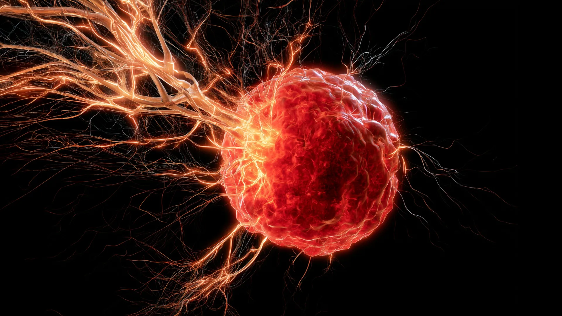
New research emanating from the University of Oklahoma has shed critical light on a previously opaque mechanism by which an aggressive form of breast cancer, specifically triple-negative breast cancer (TNBC), manipulates the body’s immune system. The groundbreaking study reveals how TNBC tumors coerce immune cells to draw nerve fibers into their mass, creating a microenvironment that appears to significantly foster cancer growth and potentially contribute to its notorious resistance to treatment. This discovery offers a novel understanding of tumor innervation and opens promising avenues for innovative therapeutic strategies.
Understanding the Aggressiveness of Triple-Negative Breast Cancer
Triple-negative breast cancer stands as one of the most challenging subtypes of breast cancer to treat. Unlike other forms, TNBC cells lack receptors for estrogen, progesterone, and the HER2 protein. This absence means that many of the highly effective targeted therapies available for other breast cancers, which work by blocking these receptors, are ineffective against TNBC. Consequently, treatment options are primarily limited to chemotherapy, surgery, and radiation, with recent advancements including immunotherapy for a subset of patients.
TNBC accounts for approximately 10-15% of all breast cancers, but its impact is disproportionately severe. It is often diagnosed in younger women, women of African ancestry, and those with BRCA1 gene mutations. Characterized by higher rates of recurrence, metastasis, and poorer overall survival compared to other breast cancer types, TNBC presents an urgent need for new therapeutic targets. The aggressive nature of TNBC underscores why understanding its underlying biological mechanisms, particularly how it interacts with its surrounding microenvironment, is so crucial for improving patient outcomes.
The Enigma of Tumor Innervation
For decades, scientists have observed that many solid tumors, including those of the breast, pancreas, and prostate, are not merely clumps of cancerous cells but intricate ecosystems containing extensive networks of blood vessels, immune cells, and notably, nerves. The presence of these nerve fibers within tumors has long been recognized, but the precise mechanisms by which they infiltrate the tumor mass and, more importantly, their functional role in cancer progression have remained largely unclear. Early hypotheses suggested that nerves might simply be bystanders, growing into the tumor incidentally. However, a growing body of evidence indicates that nerves are active participants in the tumor microenvironment, capable of influencing tumor growth, angiogenesis (the formation of new blood vessels), metastasis, and even immune evasion. The University of Oklahoma study, published in the esteemed journal Cell Death & Differentiation, provides a significant piece of the puzzle, offering a detailed explanation for how these nerves are actively recruited into TNBC tumors.
A Molecular Mechanism Unveiled: The Role of Macrophages and BDNF
The core of the OU research lies in identifying a sophisticated mechanism through which TNBC manipulates the immune system. The researchers pinpointed macrophages, a type of immune cell that normally plays a beneficial role in the body. Macrophages are the "big eaters" of the immune system, responsible for engulfing pathogens, clearing cellular debris, and initiating tissue repair. They are highly plastic cells, capable of adopting different functional states, often categorized as M1 (pro-inflammatory, anti-tumor) or M2 (anti-inflammatory, pro-tumor). In the context of cancer, tumors often reprogram macrophages to adopt an M2-like phenotype, transforming them from potential immune defenders into unwitting accomplices that support tumor growth and metastasis.
The OU team discovered that TNBC tumors actively attract these macrophages. Once within the tumor microenvironment, these recruited macrophages undergo a critical transformation. They begin to secrete a protein called brain-derived neurotrophic factor, or BDNF. BDNF is widely known for its vital role in the central nervous system, where it supports the growth, differentiation, and survival of neurons. It’s a key player in brain development, learning, and memory. However, in a stark example of cancer’s ability to hijack normal biological processes, the researchers found that TNBC tumors exploit this same neurotrophic signal. By inducing macrophages to release BDNF, the tumors create a potent chemotactic gradient that actively encourages nearby peripheral nerves to grow directly towards and penetrate the cancerous mass.
Dr. Maureen Cox, Ph.D., an assistant professor in the Department of Microbiology and Immunology at the OU College of Medicine and a research member of OU Health Stephenson Cancer Center, elaborated on this critical finding. "Macrophages are the critical source for drawing nerves into the tumor," Dr. Cox stated. "Although macrophages typically play a positive role in the body, they are facilitating a negative function in this scenario of breast cancer." This statement underscores the cunning adaptability of cancer cells, turning the body’s own defense mechanisms against it to create conditions favorable for its own survival and proliferation.
Experimental Validation and Therapeutic Promise
To validate their findings and explore potential therapeutic interventions, Dr. Cox and her colleagues moved from observation to intervention. They conducted a series of experiments using mouse models of triple-negative breast cancer. In these models, they confirmed that the presence of tumor-associated macrophages was indeed linked to increased nerve infiltration. The pivotal step involved testing a pharmacological intervention. The researchers utilized a drug that specifically blocks BDNF signaling. The results were compelling: when mice were treated with this BDNF-blocking agent, the growth of nerves into the tumors was significantly inhibited. Crucially, this reduction in nerve innervation correlated with a substantial decrease in overall tumor growth.
This finding carries immense therapeutic potential, particularly because the drug used to block BDNF signaling is already available on the market, albeit for other conditions. The prospect of repurposing an existing drug could drastically accelerate its journey to clinical trials for cancer patients. Dr. Cox expressed optimism regarding this aspect: "It looks really promising that we can use this drug, which is already on the market, to target BDNF."
Beyond merely slowing tumor growth, the researchers hypothesize a broader benefit. "We believe that the nerves are immunosuppressive," Dr. Cox explained. "So if we can stop the nerves from growing in the first place, maybe we can boost the immune response to help fight the cancer." This suggests that targeting nerve infiltration might not only directly impede tumor progression but also indirectly enhance the efficacy of immunotherapies, which aim to unleash the body’s own immune system against cancer cells. This potential synergy could be particularly impactful for TNBC patients, for whom current immunotherapies are effective in only a subset.
Translational Relevance: Evidence in Human Patients
A critical step in any preclinical research is to determine its relevance to human disease. The OU team extended their investigation by examining clinical data from human patients diagnosed with triple-negative breast cancer. Their analysis revealed a sobering correlation: tumors containing higher levels of both macrophages and BDNF were statistically linked with poorer survival rates among these patients. This direct evidence from human pathology strongly supports the translational significance of the mechanisms observed in mouse models. It suggests that the macrophage-BDNF-nerve axis is not merely an experimental curiosity but a clinically relevant pathway contributing to the aggressive nature and poor prognosis of TNBC in people. This human data provides a robust foundation for pursuing clinical trials targeting this pathway.
Broader Implications and Future Directions
The University of Oklahoma’s discovery has far-reaching implications, potentially reshaping our understanding of the tumor microenvironment and informing future therapeutic strategies across oncology. The research points towards several key areas for future investigation and development:
- Novel Therapeutic Targets: The BDNF signaling pathway emerges as a promising new target for TNBC, potentially leading to the development of novel drugs or the repurposing of existing ones. This could be particularly valuable in combination therapies, where BDNF blockers might enhance the effectiveness of chemotherapy, radiation, or immunotherapy.
- Diagnostic and Prognostic Biomarkers: The presence and activity levels of macrophages and BDNF within TNBC tumors could serve as new diagnostic or prognostic biomarkers, helping clinicians to better stratify patients, predict disease aggressiveness, and tailor treatment plans.
- Understanding Nerve-Cancer Crosstalk: The study opens the door to a deeper understanding of exactly how nerves contribute to tumor growth and metastasis. Dr. Cox and her team are eager to explore these mechanisms further. Some evidence suggests that nerves may actively stimulate the formation of new blood vessels (angiogenesis), which are essential for supplying tumors with oxygen and nutrients, thus fueling their growth. Other research indicates that cancer cells might use nerve fibers as "highways" to migrate away from the primary tumor, facilitating metastasis to distant sites. Interrupting these nerve-guided migratory pathways could be a powerful strategy to prevent the spread of cancer.
- Application to Other Cancers: The researchers are already planning to investigate the same intervention in other aggressive cancers that are notoriously difficult to treat, such as high-grade ovarian cancer. If the macrophage-BDNF-nerve axis proves to be a conserved mechanism across multiple aggressive tumor types, the therapeutic implications could be even more profound.
Ultimately, the overarching goal of this research is to leverage these insights to re-engage the body’s natural defenses. "Ultimately, we want to turn the anti-tumor immunity back on in cancer patients so their own immune systems can reject the tumors," Dr. Cox affirmed. This vision aligns with the broader paradigm shift in cancer research towards harnessing immunotherapy, aiming to empower the patient’s own body to fight the disease effectively.
A Collaborative Effort for Advancement
This significant research was made possible through robust support from various institutions. The National Institute of General Medical Sciences of the NIH (award numbers P20GM103447 and P20GM103639) provided critical funding. Additionally, Oklahoma’s Tobacco Settlement Endowment Trust (TSET), a primary funder of the Stephenson Cancer Center and TSET Health Promotion Research Center at the University of Oklahoma, played a vital role. Further support came from the Oklahoma Shared Clinical and Translational Resources through an Institutional Development Award from the National Institute of General Medical Sciences (grant no. U54GM104938). This collaborative funding model underscores the multi-faceted nature of modern scientific inquiry and the collective effort required to advance our understanding and treatment of complex diseases like cancer. The findings from the University of Oklahoma represent a substantial stride forward in the ongoing battle against triple-negative breast cancer, offering renewed hope for patients and paving the way for a new generation of targeted therapies.
