
 By Lina carolina
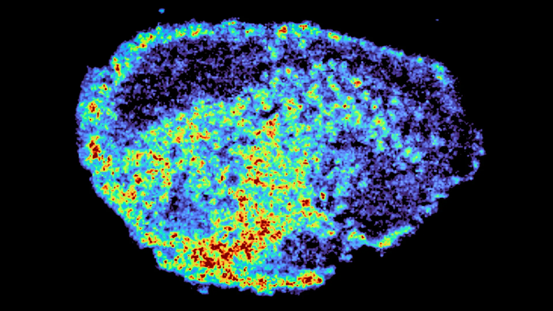
The pursuit of more effective cancer therapies is a relentless endeavor, often hampered by a fundamental challenge: the stark variability in patient response to even the most promising treatments. A groundbreaking study, published in the esteemed journal Nature Communications, is shedding critical light on this phenomenon, particularly concerning PARP inhibitors, a class of targeted drugs revolutionizing ovarian cancer care. Led by Dr. Louise Fets at the Medical Research Council (MRC) Laboratory of Medical Sciences (LMS), the research meticulously tracked the journey of these potent drugs within human ovarian tumor samples, employing sophisticated imaging techniques to uncover hidden mechanisms that dictate treatment success or failure.
The study’s pivotal finding centers on the unexpected role of lysosomes, often described as the cell’s "recycling centers." Researchers have discovered that PARP inhibitors can accumulate within these cellular organelles, becoming trapped and subsequently released. This dynamic process profoundly influences the drug’s availability within cancer cells, ultimately impacting the efficacy of the treatment. This revelation offers a crucial piece of the puzzle in understanding why a drug that performs exceptionally for one patient may prove ineffective for another, a conundrum that has long plagued oncologists and researchers alike.
The Complex Landscape of Cancer Drug Distribution
The landscape of cancer treatment has witnessed unprecedented advancements in recent years, offering renewed hope and improved prognoses for a growing number of patients. Among these innovations, PARP inhibitors have emerged as transformative agents, particularly in the management of ovarian cancer. These drugs function by targeting a specific weakness in cancer cells that have defects in DNA repair pathways, leading to their selective destruction. However, the clinical reality is that not all patients derive the same benefit, and the development of resistance over time remains a significant hurdle.
For PARP inhibitors, like many targeted therapies, to exert their life-saving effects, they must achieve and maintain sufficiently high concentrations within cancer cells to trigger programmed cell death (apoptosis). Despite this fundamental requirement, the intricate processes governing drug distribution within the complex architecture of tumors, and crucially, within individual cancer cells, have remained largely elusive. This lack of granular understanding has limited the ability to optimize drug delivery and predict treatment responses with precision.
The research conducted by Dr. Fets and her team addresses this knowledge gap directly. By utilizing patient-derived ovarian tumor explants – meticulously preserved, living tumor tissue maintained outside the body – the scientists were able to observe drug behavior in a setting that closely mimics the in vivo environment. These explants were treated with PARP inhibitors, allowing for direct visualization of drug movement through authentic human tumor tissue. This innovative approach moves beyond cell line studies, providing a more relevant and clinically applicable understanding of drug dynamics.
The team employed a powerful combination of advanced analytical tools. Mass spectrometry imaging was instrumental in generating detailed, high-resolution maps that pinpointed the precise locations of drug accumulation within the tumor samples. This was synergistically paired with spatial transcriptomics, a cutting-edge technology that allows for the examination of gene expression patterns within specific spatial regions of a tissue sample. By overlaying these datasets, researchers could correlate areas of high and low drug concentration with the underlying genetic activity of the cancer cells. The results were striking, revealing significant heterogeneity in drug distribution, not only between different tumors from different patients but also within individual tumor samples, even when subjected to identical drug dosages.
"A novel aspect of this study was the use of mass spectrometry imaging to directly measure and visualize drug uptake in patient tumour tissue," explained Dr. Zoe Hall, senior author of the study and Associate Professor at Imperial College London’s Department of Metabolism, Digestion and Reproduction. "Through the spatial mapping of drug molecules, we could pinpoint regions of high and low drug and compare gene expression, from the same tissue slice, using spatial transcriptomics." This dual-pronged approach provided an unprecedentedly detailed view of the drug’s journey, laying the foundation for understanding the observed variations in therapeutic outcomes.
Lysosomes: Unveiling the Hidden Drug Reservoirs
The most significant revelation from the study is the central role that lysosomes play in this observed heterogeneity of drug distribution. Lysosomes, membrane-bound organelles within cells, are primarily responsible for breaking down waste materials and cellular debris. However, this research demonstrates that certain PARP inhibitors are actively taken up by lysosomes and sequestered within these compartments, rather than distributing uniformly throughout the cell. This creates localized pockets of high drug concentration within the cell, while other regions may experience significantly lower levels.
These lysosomes, therefore, function as sophisticated, albeit unintended, drug reservoirs. They can hold onto the PARP inhibitors, releasing them gradually over time. This slow-release mechanism leads to prolonged exposure in some cellular compartments and cells, potentially enhancing therapeutic effects, while others may be exposed to insufficient drug levels to achieve the desired cell death. The study also highlighted that not all PARP inhibitors are equally susceptible to this lysosomal sequestration. Specifically, drugs such as rucaparib and niraparib were found to be significantly influenced by this lysosomal accumulation, whereas olaparib, another widely used PARP inhibitor, did not exhibit the same behavior. This differential interaction suggests that the chemical properties of the drugs themselves play a critical role in their cellular trafficking and subsequent accumulation.
"We were surprised to see large variability in drug accumulation at the single-cell level," stated Dr. Carmen Ramirez Moncayo, the study’s first author and a Postdoctoral Researcher at the LMS. "This variability was driven by the build-up of a drug in lysosomes, which are acting as reservoirs, increasing the exposure of cancer cells to drugs, by storing and releasing the drug when needed." This observation underscores the complexity of drug-cell interactions and the need to consider intracellular dynamics when evaluating drug efficacy.
Implications for the Future of Personalized Cancer Treatment
The implications of this research are far-reaching, particularly in the context of optimizing current cancer therapies and developing future treatment strategies. PARP inhibitors are not only vital in ovarian cancer but are also approved for use in breast and prostate cancers, with ongoing clinical trials exploring their potential in a growing array of other cancer types. A deeper understanding of how these drugs are stored and distributed within cells could pave the way for more personalized treatment approaches. This could involve tailoring drug selection based on a patient’s specific tumor biology, optimizing dosages and schedules to maximize drug exposure in critical cellular compartments, and potentially mitigating the development of drug resistance and subsequent relapses.
"By understanding how drugs are taken up into cells, we can understand whether this influences why cancer drugs work for some people and not for others," Dr. Louise Fets, senior author and Head of the LMS’ Drug Transport and Tumour Metabolism Group, emphasized. "Eventually, we hope to be able study the molecular signature of a patient’s tumor to help to tailor therapeutic approaches in a more personalized way." This vision of precision oncology, where treatments are meticulously designed for the individual patient, is a key aspiration of modern cancer research.
It is crucial to acknowledge the limitations of the current study, which utilized tumor tissue maintained ex vivo. In a living patient, drug delivery is primarily through the bloodstream. Furthermore, tumor vasculature is often characterized by disorganization and abnormalities, which can further exacerbate uneven drug distribution. These factors, coupled with the newly identified lysosomal sequestration, create a complex interplay that likely contributes to treatment variability.
Future research will undoubtedly build upon these findings. The next steps will involve employing animal models and expanding studies to larger patient cohorts to more comprehensively investigate how drug delivery mechanisms, tumor microenvironment characteristics, and the newly elucidated lysosomal storage dynamics interact within clinical settings. This includes a particular focus on understanding drug behavior in relapsed cancers, where resistance mechanisms are often more pronounced.
The research was made possible through substantial funding from a consortium of esteemed organizations, including the Medical Research Council, Cancer Research UK, a PhD studentship from the Integrative Toxicology Training Partnership administered by the MRC Toxicology Unit, and a Victoria’s Secret Global Fund for Women’s Cancers Career Development Award, in partnership with Pelotonia and the American Association for Cancer Research (AACR). This collaborative effort highlights the significant investment and commitment towards unraveling the complexities of cancer and improving patient outcomes.
In essence, this study represents a significant leap forward in our comprehension of how cancer drugs operate at the cellular level. By illuminating the critical role of lysosomes as drug reservoirs, the research provides a tangible explanation for treatment variability and opens new avenues for developing more intelligent, personalized, and ultimately, more effective cancer therapies. The journey from laboratory discovery to clinical application is often long, but this work offers a compelling roadmap for a future where cancer treatment is more precisely tailored to each individual’s unique biological landscape.
