
 By Nana O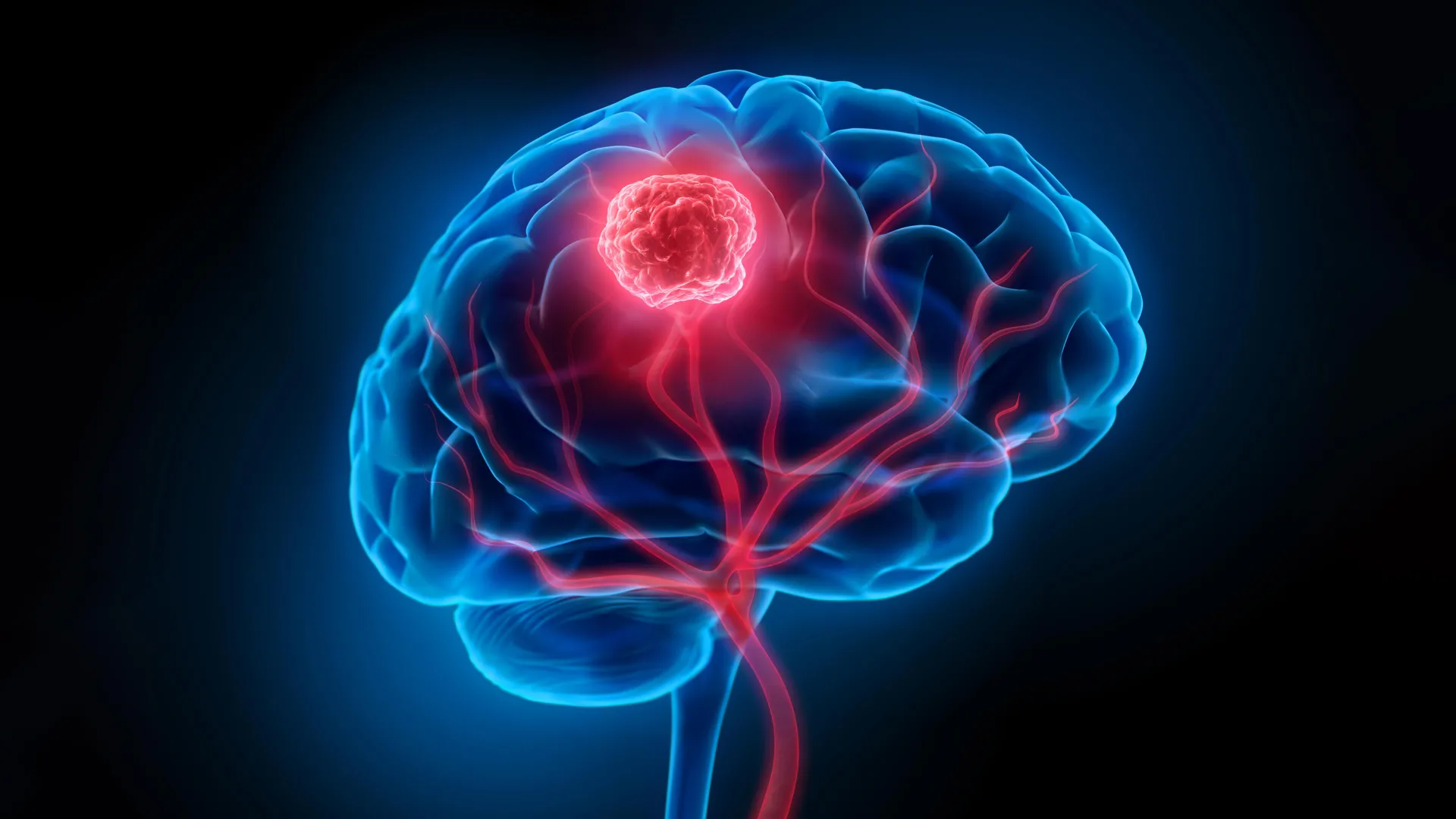
Glioblastoma, a notoriously aggressive form of brain cancer, has long presented a formidable challenge to the medical community, largely due to its invasive nature and the intricate complexities of the brain environment. The disease’s relentless spread into surrounding healthy brain tissue makes complete surgical resection a precarious endeavor, requiring surgeons to navigate a delicate balance between tumor removal and the preservation of vital neurological function. Compounding this surgical hurdle is the formidable blood-brain barrier, a biological shield that severely limits the efficacy of conventional treatments like chemotherapy and radiotherapy, preventing them from adequately reaching and eradicating tumor cells. These intertwined obstacles contribute significantly to the grim prognosis for glioblastoma patients, with the five-year survival rate hovering at a mere 7 percent, underscoring the urgent need for innovative therapeutic strategies.
In a significant stride toward overcoming these entrenched difficulties, researchers from the University of Technology Sydney (UTS), in collaboration with Harvard University and Henan University, have unveiled a groundbreaking ‘double-punch’ nanozyme platform. This pioneering system, detailed in a recent publication in the esteemed journal Science Translational Medicine, utilizes a single, intelligent nanoparticle design to simultaneously address both the surgical visualization and post-operative treatment challenges inherent in glioblastoma management.
A Precisely Engineered Dual-Function Nanomaterial
At the heart of this innovative platform lies a meticulously engineered, two-dimensional atomic sheet, constructed with atomic precision through a process adapted from advanced semiconductor manufacturing techniques. This unique material architecture empowers the nanoparticle to seamlessly transition between two critical roles, acting as both a high-precision surgical guide and a targeted therapeutic agent. The dual functionality is activated by a single, non-invasive source: near-infrared light.
Dr. Bingyang Shi, Chair Professor of Nanomedicine at UTS’s School of Electrical, Mechanical and Biomedical Engineering and a lead author on the study, elaborated on the transformative potential of this integrated system. "We’ve engineered a single material that does two jobs in sequence," Professor Shi explained. "It’s a precise guide for the surgeon during the operation, and then a targeted clean-up treatment afterward." This sequential application, driven by a single activation mechanism, streamlines the treatment process and minimizes the complexity for both clinicians and patients.
Enhancing Surgical Visualization for Unprecedented Precision
During surgical intervention, the nanozyme platform functions as an exceptionally sensitive imaging agent. A specially engineered fluorescent dye integrated onto the atomic sheet emits a glow when exposed to near-infrared wavelengths, a spectrum invisible to the human eye. This targeted fluorescence allows surgeons to discern individual tumor cell clusters with remarkable clarity, even those as small as 44 micrometers – a level of resolution that surpasses the capabilities of current clinical imaging technologies. Furthermore, the nanoparticle is equipped with a targeting molecule designed to facilitate its passage across the blood-brain barrier and ensure its specific accumulation within glioma cells, thereby maximizing its visibility in the tumor microenvironment.
The ability to visualize these microscopic tumor extensions is a critical advancement. Glioblastoma tumors are notoriously diffuse, often infiltrating surrounding brain tissue in a way that is difficult to demarcate precisely. Traditional imaging techniques, while valuable, may not always detect the smallest infiltrations, leaving surgeons to rely on their expertise and visual cues that can be fallible when dealing with such insidious cellular spread. The enhanced visualization provided by this nanoparticle platform promises to significantly improve the accuracy and completeness of tumor resection.
Targeted Post-Operative Therapy to Eliminate Residual Disease
Following the visible tumor’s removal, the same nanoparticle material can be reintroduced into the surgical cavity and re-activated using the same near-infrared light. This triggers its therapeutic function, a process termed phototherapy. In this phase, the platinum atoms embedded within the nanoparticle play a crucial role. They catalyze a reaction that converts the tumor’s own hydrogen peroxide into oxygen. This localized oxygen production is vital, as glioblastoma cells often thrive in hypoxic (low-oxygen) environments, which can shield them from conventional treatments. By increasing oxygen levels, the therapy creates a less hospitable environment for any remaining cancer cells.
Simultaneously, the near-infrared light activation generates heat and reactive oxygen species (ROS). These localized effects are designed to destroy any microscopic cancer cells that may have evaded surgical removal, cells that are often responsible for tumor recurrence. This two-pronged therapeutic approach – oxygen generation and localized thermal and chemical assault – offers a potent strategy for eradicating residual disease that is otherwise undetectable and untreatable by conventional means.
Addressing the Critical Challenge of Tumor Recurrence
The persistence of microscopic cancer cells after surgery is a primary driver of glioblastoma recurrence, a devastating outcome for patients. Even if the bulk of the tumor is successfully excised, a few stray cells can initiate new tumor growth, often leading to a more aggressive and treatment-resistant disease. The nanozyme platform is specifically engineered to combat this critical issue.
Pre-Clinical Success in Animal Models
The efficacy of this dual-action nanoparticle platform has been rigorously tested in preclinical studies utilizing mouse models of glioblastoma. The results have been highly encouraging. In these studies, mice treated with the nanoparticle approach following surgery demonstrated a significant reduction in tumor recurrence. Crucially, all treated mice survived for at least 60 days post-treatment, a marked improvement compared to the 42-day survival observed in mice that underwent surgery alone. Furthermore, extensive follow-up testing revealed no detectable neurological or motor impairments in the treated animals, suggesting a favorable safety profile in this preclinical context.
The implications of these findings are substantial. If replicated in human trials, this technology could dramatically alter the treatment paradigm for glioblastoma, moving beyond solely debulking the visible tumor to actively eliminating the microscopic threat that fuels its return.
A Promising Avenue, Yet Early-Stage Research
While the results are undeniably promising, the research team emphasizes the preliminary nature of these findings. "The results are very encouraging, but this is still early-stage research carried out in mouse models, not in people – and that distinction is important," Professor Shi cautioned. The transition from animal models to human application involves a complex and rigorous series of clinical trials. The scale and complexity of the human brain present unique challenges, and the imaging and therapeutic performance of the nanoparticle will need to be validated at this larger scale.
Broader Implications and Future Outlook
The development of this dual-function nanozyme platform represents a significant conceptual leap in the fight against glioblastoma. By integrating diagnostic and therapeutic capabilities into a single, sophisticated nanomaterial, researchers are paving the way for more precise and effective cancer treatments. The potential for surgeons to visualize more of the tumor during operations and for a targeted therapy to eliminate residual disease afterward offers a tangible hope for reducing recurrence rates, a goal that has eluded medical professionals for decades.
The broader impact of this research extends beyond glioblastoma. The principles of creating tunable, multi-functional nanomaterials that can navigate biological barriers and perform sequential tasks could be applied to the treatment of other complex cancers or diseases affecting the central nervous system.
The timeline for translating this laboratory breakthrough into a clinical reality is yet to be determined and will depend on the successful navigation of rigorous human clinical trials, regulatory approvals, and manufacturing scalability. However, the scientific community will be closely watching the progress of this innovative technology. The potential to equip surgeons with enhanced visualization tools and to provide a potent post-operative therapy offers a beacon of hope for patients facing this devastating diagnosis. If this technology continues to prove its efficacy and safety through further research and clinical validation, it could herald a new era in the management of glioblastoma, offering a meaningful step towards improving outcomes and extending the lives of those affected by this aggressive brain cancer. The collaborative effort between institutions like UTS, Harvard, and Henan universities highlights the power of international scientific cooperation in tackling some of the world’s most pressing medical challenges.
