
 By George Ongkojoyo
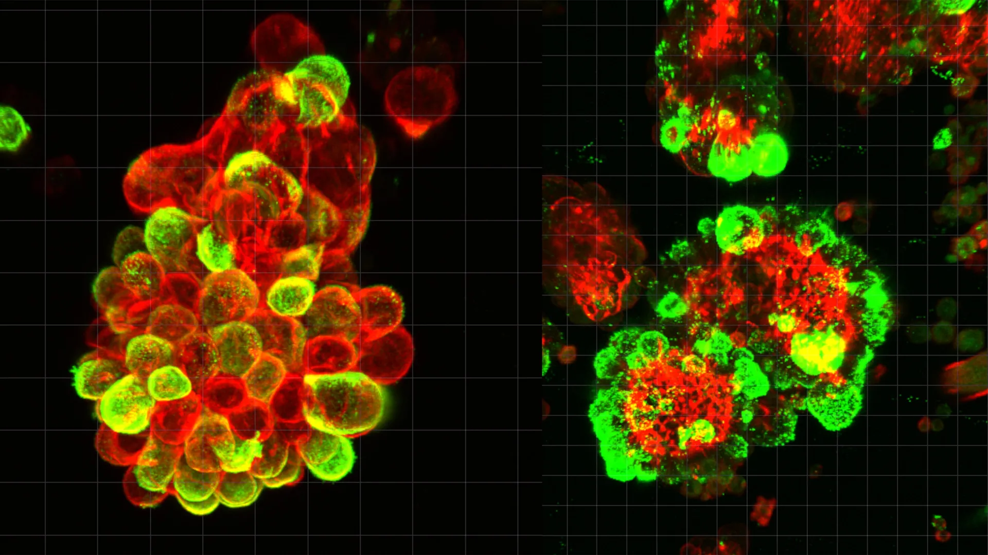
Ovarian cancer remains the most lethal of all gynecological malignancies, characterized by a silent progression that often evades detection until it has reached advanced stages. For decades, the medical community has grappled with the rapid and aggressive nature of this disease, which frequently presents as widespread abdominal tumors before a primary diagnosis is even established. A groundbreaking study led by researchers at Nagoya University, recently published in the journal Science Advances, has finally provided a mechanical explanation for this phenomenon. The research identifies a sophisticated "cooperation" between malignant cells and healthy mesothelial cells—the very cells meant to protect the abdominal cavity—revealing that ovarian cancer does not spread in isolation but rather through the formation of highly invasive, hybrid cell clusters.
The Silent Killer and the Mystery of Rapid Spread
Ovarian cancer is often referred to by oncologists as the "silent killer" because its early symptoms, such as bloating and pelvic discomfort, are frequently mistaken for benign digestive issues. According to the American Cancer Society, when ovarian cancer is diagnosed at a localized stage, the five-year survival rate is approximately 93%. However, only about 15% of cases are found this early. The vast majority are diagnosed at Stage III or IV, where the cancer has already disseminated throughout the peritoneal cavity, dropping the survival rate significantly.
The central mystery has always been the speed of this dissemination. Unlike many other solid tumors that spread through the lymphatic system or the bloodstream, ovarian cancer utilizes the fluid within the abdominal cavity. While researchers understood the route, the biological drivers that allowed these cells to attach to and penetrate distant organs so efficiently remained poorly understood. The Nagoya University study marks a paradigm shift in this understanding by demonstrating that cancer cells effectively "outsource" the mechanical work of invasion to non-cancerous cells.
The Discovery of Hybrid Cell Clusters
The research team, led by Dr. Kaname Uno and colleagues at the Nagoya University Graduate School of Medicine, began their investigation by analyzing the abdominal fluid, or ascites, of patients diagnosed with ovarian cancer. Using high-resolution microscopy and single-cell RNA sequencing, they observed a phenomenon that challenged the traditional view of metastatic cancer cells as solitary travelers.
Rather than drifting as individual units, the cancer cells were found to be part of compact, spherical clusters. Crucially, these spheres were not composed solely of malignant cells. In approximately 60% of the observed clusters, the cancer cells had recruited mesothelial cells—specialized cells that normally form a smooth, protective lining (the mesothelium) over the internal organs and the abdominal wall.
The study revealed that this is not a random association. Ovarian cancer cells actively secrete a signaling protein known as Transforming Growth Factor-beta 1 (TGF-β1). This molecule acts as a chemical command, hijacking the biological programming of the recruited mesothelial cells. Under the influence of TGF-β1, the mesothelial cells undergo a transformation, changing from passive lining cells into active, aggressive "pathfinders" for the tumor.
Invadopodia: The Biological Drill Bits
The most striking finding of the Nagoya University study involves the mechanical changes that occur within these hybrid clusters. Once the mesothelial cells are integrated into the sphere and activated by TGF-β1, they begin to develop invadopodia. These are specialized, spike-like protrusions rich in actin and proteolytic enzymes.
In a healthy context, mesothelial cells provide a frictionless surface for organs to move against one another. However, in the hybrid cluster, the mesothelial cells use their newly formed invadopodia to act as biological drill bits. As the cluster floats through the abdominal fluid and eventually makes contact with an organ—such as the liver, stomach, or intestinal wall—the mesothelial cells lead the charge. They use their invadopodia to pierce the protective surface of the target organ, creating microscopic openings.
Dr. Kaname Uno, the study’s lead author, noted that the cancer cells themselves remain relatively passive during this initial breach. "They manipulate mesothelial cells to do the tissue invasion work," Dr. Uno explained. "The cancer cells undergo minimal genetic and molecular changes during this specific phase; they simply migrate through the openings that the mesothelial cells have already created." This "Trojan Horse" strategy allows the cancer to spread with far less energy and genetic adaptation than previously thought.
A Divergent Path: Ovarian vs. Other Cancers
The research highlights a fundamental difference in how ovarian cancer spreads compared to other common cancers like breast or lung cancer. In those diseases, metastasis typically involves "hematogenous spread," where cancer cells intravasate into blood vessels and travel through the circulatory system. Because blood follows a predictable, pressurized circuit, clinicians can often use "liquid biopsies" to detect circulating tumor cells (CTCs) or cell-free DNA to monitor the disease.
Ovarian cancer, however, largely bypasses the bloodstream. It spreads via "peritoneal dissemination," where cells break off from the primary tumor and enter the peritoneal fluid. This fluid is in constant, unpredictable motion, driven by the rhythmic movements of the diaphragm during breathing and the peristaltic waves of the digestive tract. This lack of a fixed route, combined with the formation of hybrid clusters that shield the cancer cells, explains why traditional blood tests often fail to catch the early stages of the spread. The Nagoya University findings suggest that the "floating phase" in the abdominal fluid is the most critical window for the cancer’s transition from a localized tumor to a systemic disease.
Resistance to Chemotherapy
Beyond their ability to invade tissue, these hybrid cell clusters offer the cancer a significant survival advantage against modern medicine. The study found that these spheres are markedly more resistant to chemotherapy than individual cancer cells or clusters composed only of malignant cells.
There are two primary reasons for this resistance. First, the physical structure of the sphere, with mesothelial cells integrated into the architecture, creates a dense barrier that limits the penetration of cytotoxic drugs like carboplatin and paclitaxel. Second, the biological signaling between the mesothelial cells and the cancer cells appears to trigger survival pathways that protect the malignant cells from apoptosis (programmed cell death) typically induced by chemotherapy. This finding provides a potential explanation for why many ovarian cancer patients initially respond well to treatment, only to suffer a recurrence of more aggressive, chemo-resistant tumors later.
From the Clinic to the Laboratory: Dr. Kaname Uno’s Motivation
The impetus for this research was deeply personal for the lead author. Before transitioning into full-time research, Dr. Kaname Uno spent eight years as a practicing gynecologist. During his time in the clinic, he encountered a patient whose case would define his career trajectory.
The patient had undergone a routine gynecological screening and received a completely normal result. However, only three months later, she returned with severe symptoms and was diagnosed with advanced-stage ovarian cancer that had already spread throughout her abdomen. Despite the best available treatments, the disease progressed too rapidly to save her life.
"That experience stayed with me," Dr. Uno remarked. "The fact that our existing diagnostic tools could miss a disease that would become terminal in just ninety days suggested there was a fundamental gap in our understanding of how this cancer moves." This motivation led him to Nagoya University’s Graduate School of Medicine, where he sought to uncover the cellular mechanics of that rapid progression.
Methodology and Validation
To ensure the robustness of their findings, the Nagoya University team employed a multi-faceted experimental approach. They utilized:
- Patient-Derived Samples: Direct analysis of ascites fluid from patients provided "real-world" evidence of the hybrid clusters.
- Advanced Microscopy: Time-lapse imaging allowed the researchers to watch the mesothelial cells forming invadopodia and drilling into substrates in real-time.
- Mouse Models: By injecting hybrid clusters into the abdominal cavities of mice, the researchers confirmed that these clusters led to more rapid and widespread tumor formation compared to injections of cancer cells alone.
- Single-Cell RNA Sequencing: This allowed the team to map the gene expression profiles of the cells within the clusters, confirming the TGF-β1 signaling pathway as the primary driver of the mesothelial transformation.
Implications for Future Treatment and Diagnostics
The identification of mesothelial cells as "unwitting accomplices" in ovarian cancer spread opens several new avenues for therapy. Current treatment protocols almost exclusively target the cancer cells themselves. This research suggests that a dual-target approach may be necessary.
One potential strategy involves the development of drugs that block the TGF-β1 signaling pathway. By preventing the cancer cells from "recruiting" the mesothelial cells, doctors might be able to keep the cancer cells in a less invasive, solitary state, making them more vulnerable to standard chemotherapy. Another possibility is the development of therapies that specifically target the formation of invadopodia, effectively "disarming" the clusters before they can attach to new organs.
Furthermore, the study suggests a new diagnostic frontier. If these hybrid clusters can be reliably identified and quantified in abdominal fluid, it could lead to the development of a "peritoneal biopsy" that is far more accurate than current blood-based markers like CA-125. Monitoring the presence of these clusters could help oncologists predict which patients are at the highest risk for rapid metastasis and allow for earlier, more aggressive intervention.
Conclusion
The Nagoya University study provides a long-overdue explanation for the aggressive nature of ovarian cancer. By demonstrating that the disease’s speed is not just a function of the cancer cells themselves, but of their ability to hijack and transform the body’s own protective cells, the research offers a clearer roadmap for future intervention. As the medical community moves toward personalized and targeted medicine, understanding these cellular partnerships will be essential in turning the tide against one of the most challenging diseases in women’s health. The work of Dr. Uno and his team underscores the vital link between clinical observation and laboratory discovery, offering hope that the "silent killer" may one day be silenced by science.
