
 By Nana O
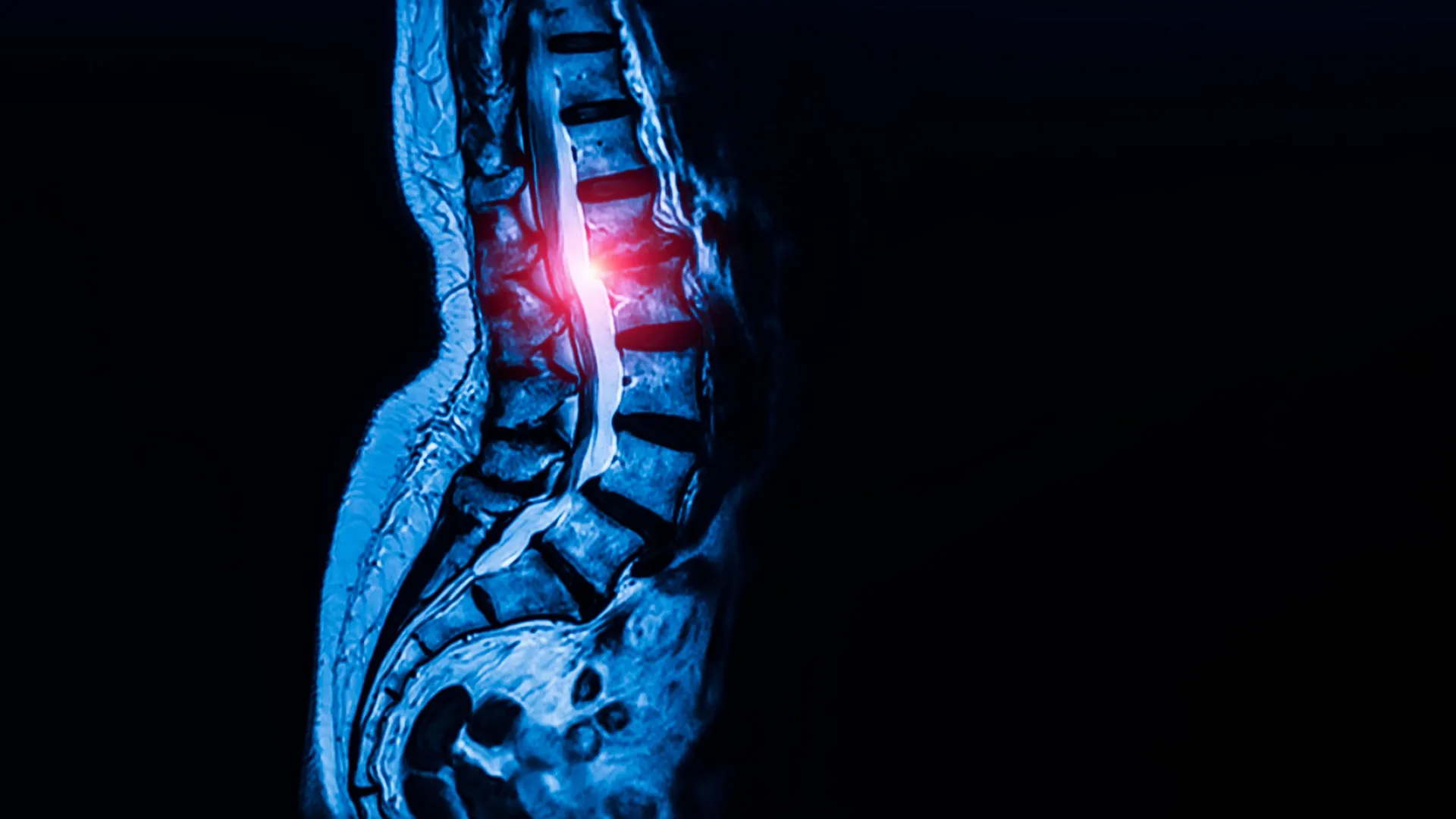
Researchers at Weill Cornell Medicine and the Hospital for Special Surgery (HSS) have achieved a groundbreaking discovery: the identification of a previously elusive stem cell population that appears to be the sole source of the body’s tendons and ligaments, the vital connective tissues that anchor muscles to bones and stabilize joints. This seminal finding, published on September 7th in the prestigious journal Cell, not only sheds light on fundamental principles of human musculoskeletal development but also opens promising new avenues for treating debilitating conditions like lumbar spinal stenosis, which affects an estimated 103 million individuals worldwide.
For years, the precise cellular origins of tendons and ligaments have remained a significant enigma in regenerative medicine and developmental biology. While various candidate stem cell populations have been proposed, none had definitively demonstrated the capacity for both self-renewal and the generation of the diverse array of mature cell types that constitute these crucial connective tissues. This new research, spearheaded by co-corresponding authors Dr. Matthew Greenblatt, a pathologist at Weill Cornell Medicine and NewYork-Presbyterian/Weill Cornell Medical Center, and Dr. Sravisht Iyer, an orthopedic professor at Weill Cornell and a spine surgeon at HSS, definitively pinpoints this critical progenitor cell.
"While previous studies had proposed several candidate stem cells, none had definitively shown that a single cell population could both self-renew and generate the full spectrum of tendon and ligament cell types," explained Dr. Greenblatt, who also holds the Rohr Family Research Scholar title and is an associate professor of pathology and laboratory medicine at Weill Cornell. "This discovery is transformative because it provides a clear cellular blueprint for how these essential tissues are formed and maintained throughout life."
The significance of this discovery extends beyond fundamental science. The research team also found compelling evidence that these newly identified stem cells can become abnormally overactive in the lower spine, a phenomenon that may directly contribute to the development of lumbar spinal stenosis. This condition arises when the ligaments within the spinal canal thicken and encroach upon the space, leading to compression of spinal nerves. The consequences for patients can be severe, manifesting as chronic pain, persistent numbness, and profound difficulty with mobility, significantly impacting their quality of life.
A Decade-Long Quest for the Master Connective Tissue Cell
The journey to identify these specialized stem cells was a methodical and persistent endeavor, building upon years of prior research by Dr. Greenblatt and his team. Their previous work had successfully identified key skeletal stem cell populations involved in other crucial regenerative processes. In 2018, the group achieved a significant milestone by identifying the stem cell responsible for initiating fracture repair within the periosteum, the outer membrane of bone. Subsequently, they uncovered stem cells integral to the formation of the skull and spine, underscoring their expertise in dissecting the complex cellular choreography of skeletal development.
However, isolating the stem cells responsible for tendons and ligaments presented a unique set of challenges. Unlike bone, these connective tissues are characterized by a high density of fibroblast-like cells, which share a remarkably similar morphology. This cellular homogeneity made it exceedingly difficult to distinguish a specific progenitor population from the general cellular milieu.
"We analyzed thousands of individual cells and sorted them into individual cell types," Dr. Greenblatt elaborated on the intricate methodology. "Then we identified which one had the properties we associate with ‘stemness.’" The concept of "stemness" in this context refers to the cell’s dual capacity: its ability to replicate itself indefinitely (self-renewal) and its potential to differentiate into the specialized mature cells required for the formation and ongoing maintenance of tendon and ligament tissue.
Unveiling the "Stemness" of Tendon and Ligament Progenitors
Through meticulous analysis, the researchers identified these elusive cells residing within specialized niches in mice, located deep within tendons and ligaments. These specific microenvironments appear to function as critical reservoirs, supporting tissue growth and facilitating repair mechanisms.
The team then meticulously translated their findings from the murine model to human tissue. Human ligament samples were ethically sourced from tissue removed during surgical procedures by Dr. Iyer from consenting patients. This crucial step allowed for direct comparison and validation of their discoveries in a human context.
Dr. Lingling Hu, a postdoctoral fellow in both Dr. Greenblatt’s and Dr. Iyer’s labs, served as the first author on the study and played a pivotal role in confirming that the human cells possessed the same critical properties of self-renewal and the capacity to generate mature ligament cells. The widespread presence of these cells was also a significant finding.
"We looked in the kneecap ligament; we looked at the Achilles tendon; and everywhere we looked, we found this cell," Dr. Greenblatt stated with evident enthusiasm. "So, we think this is the universal stem cell for tendons and ligaments throughout the body." This universal nature implies that a single cellular origin is responsible for the formation of these diverse connective tissues across the entire musculoskeletal system.
The Link Between Stem Cells and Lumbar Spinal Stenosis
With the identification of the tendon and ligament stem cells firmly established, the researchers turned their attention to their potential role in lumbar spinal stenosis. They embarked on a comparative study, analyzing stem cells extracted from patients diagnosed with spinal stenosis against those collected from spinal ligaments of individuals with herniated discs but no signs of stenosis. All participating patients provided informed consent prior to their surgical procedures.
The results were striking: ligaments from individuals with spinal stenosis contained a significantly higher number of these newly identified stem cells compared to the control group. Further experiments in mice demonstrated that transplanting these "stenotic" stem cells led to the generation of more tendon cells than stem cells derived from healthy ligaments.
"Though spinal stenosis is a complex condition, this really showed us that these cells are contributing to the pathology," emphasized Dr. Greenblatt, who is also affiliated with the Sandra and Edward Meyer Cancer Center at Weill Cornell. This finding provides a direct cellular link between the overactivity of these stem cells and the thickening of ligaments characteristic of spinal stenosis.
Delving deeper into the cellular mechanisms, the researchers observed distinct differences in calcium signaling within the stem cells associated with stenosis. Calcium signaling is a fundamental cellular communication pathway that regulates various cellular processes, including growth and activity. Stem cells implicated in spinal stenosis exhibited heightened calcium signaling compared to their healthy counterparts.
A Promising Therapeutic Target: Calcium Channel Blockers
The implications of this heightened calcium signaling were profound. When the scientists experimentally increased calcium signaling in stem cells from healthy ligaments, it triggered excessive tissue growth, mimicking the pathological changes observed in spinal stenosis. Conversely, when they reduced calcium signaling in a mouse model of lumbar spinal stenosis, the abnormal cell growth was effectively inhibited.
This critical observation strongly suggests that calcium signaling pathways could serve as a potent therapeutic target for spinal stenosis. Furthermore, it opens the door to exploring the potential of calcium channel blockers – a well-established class of drugs currently prescribed for managing high blood pressure – as a novel treatment strategy for this prevalent spinal condition. While extensive clinical trials will be necessary to ascertain the safety and efficacy of this approach in human patients, the initial findings are exceptionally encouraging.
"This is probably the first work that’s shown a potential therapeutic target for one of the most common spine conditions in the world," stated Dr. Iyer, highlighting the immense clinical potential of this discovery. "The findings are exciting for their potential to change the way we deliver spinal care."
Broader Implications for Connective Tissue Disorders
The impact of this research may extend far beyond spinal stenosis. Dr. Greenblatt indicated plans to investigate whether these same stem cells are implicated in other connective tissue disorders. Conditions such as Marfan syndrome, a genetic disorder that affects connective tissues throughout the body, could potentially be addressed through a deeper understanding of these progenitor cells.
Moreover, the discovery offers a potential explanation for the often-challenging healing process of various tendon and ligament injuries. Areas ripe for future investigation include the chronic difficulties associated with rotator cuff tears, Achilles tendon injuries, and the complexities of ligament reconstruction. The research could also shed light on the underlying cellular mechanisms contributing to chronic tendon degeneration.
"Given that this cell appears to be the ultimate origin of all tendon and ligament cells, defects in this cell are likely at the heart of a wide range of tendon and ligament disorders," Dr. Greenblatt concluded. This unified perspective suggests that a single cellular target could offer therapeutic benefits across a spectrum of musculoskeletal ailments.
This groundbreaking research was supported by a multitude of esteemed funding bodies, including the Marfan Foundation’s Victor A. McKusick Fellowship, a Kellen Scholars Award, the Children’s Tumor Foundation, the Arthritis National Research Foundation, a Jumpstart award from Weill Cornell, a National Research Foundation of Korea award, the National Institutes of Health, a MIND Prize from the Pershing Square Foundation, a Mary Kay Ash Foundation Award, an Innovator Award from the Marfan Foundation, and a Burroughs Wellcome Career Award for Medical Scientists. The collaborative effort and diverse funding underscore the broad significance and potential impact of this pivotal discovery.
