
 By Gabriel Paijo
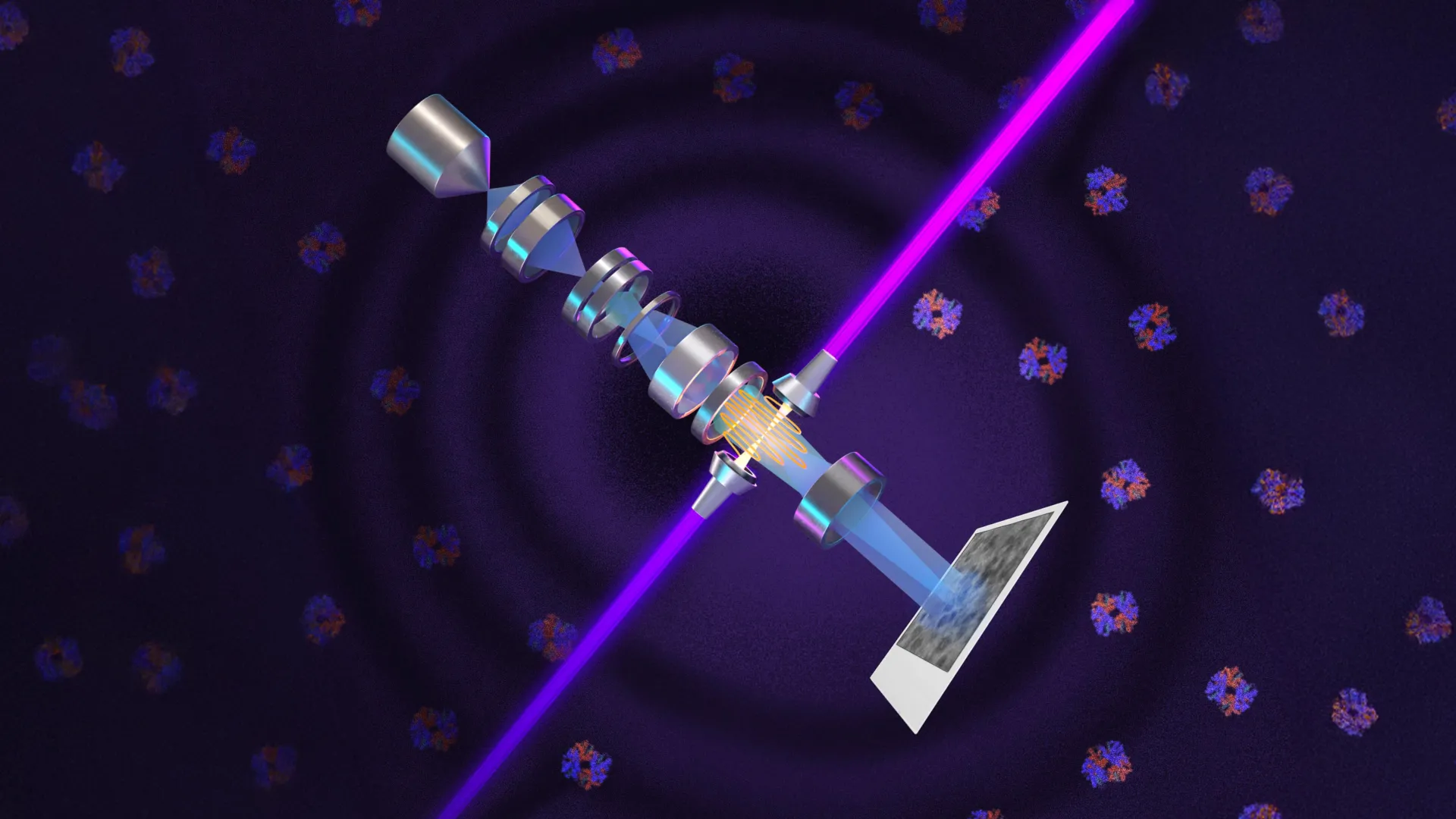
Physicists at the University of California, Berkeley, have achieved a landmark breakthrough in imaging technology by successfully integrating a high-intensity laser system into electron microscopy, a feat that promises to redefine the boundaries of biological research. By applying the principles of phase-contrast microscopy—a technique that earned the Nobel Prize in Physics over 70 years ago—to the realm of electron beams, the research team has developed a "laser phase plate" capable of magnifying cellular structures with unprecedented clarity. This advancement addresses a fundamental limitation in cryoelectron microscopy (cryo-EM), potentially opening the door to the study of the vast majority of human proteins that were previously too small to be visualized.
The development, led by Holger Müller, a UC Berkeley professor of physics and faculty scientist at Lawrence Berkeley National Laboratory, introduces a method to significantly amplify the signal-to-noise ratio in cryo-EM and its three-dimensional counterpart, cryoelectron tomography (cryo-ET). For decades, scientists have struggled to capture clear images of small proteins and complex molecular interactions within the crowded environment of a living cell. The new laser technology aims to resolve this by utilizing what researchers describe as the world’s most intense focused continuous-wave laser to alter the phase of electrons, thereby creating high-contrast images of biological specimens without the need for destructive stains or excessive radiation.
The Evolution of Phase Contrast: From Light to Electrons
To appreciate the magnitude of the UC Berkeley breakthrough, one must look back to 1930, when Dutch physicist Frits Zernike revolutionized light microscopy. At the time, biologists faced a persistent problem: living cells are mostly water and appear transparent under a standard microscope. While staining cells could provide contrast, the chemicals involved invariably killed the specimen, preventing the study of life in its active state.
Zernike discovered that as light passes through a biological structure, it does not just change in brightness; it undergoes a "phase shift." While the human eye cannot perceive these minute delays in light wave peaks, Zernike found that by shifting the unscattered light by 90 degrees, he could convert these invisible phase changes into visible differences in brightness. This discovery, known as phase-contrast microscopy, allowed scientists to see internal cellular components like the nucleus and mitochondria in living, unstained cells. The invention was so transformative that Zernike was awarded the Nobel Prize in Physics in 1953.
As electron microscopy emerged in the mid-20th century, offering magnification levels 10,000 times greater than light microscopes, scientists immediately sought to apply Zernike’s phase-contrast principle to electron beams. However, the technical hurdles were immense. Early attempts to use physical material plates to shift the phase of electrons often resulted in beam instability, loss of resolution, or total obstruction of the image. The quest for a "phase plate" for electron microscopes remained one of the great "holy grails" of imaging physics for nearly 80 years.
A 15-Year Engineering Odyssey
The solution finally began to take shape in 2010, when Holger Müller and Robert Glaeser, a pioneer in cryo-EM and UC Berkeley professor emeritus, proposed a radical idea: instead of using a physical object to shift the phase of the electron beam, they could use light. By using a focused, incredibly intense laser, they theorized they could interact with the electrons via the Ponderomotive effect, shifting their phase without the drawbacks of physical hardware.
Realizing this theory required a 15-year engineering effort. The primary challenge was the sheer amount of power required. To achieve a 90-degree phase shift in an electron beam, the researchers needed a laser focus of extraordinary intensity. Their eventual solution involved a sophisticated optical cavity—a spherical chamber lined with ultra-reflective mirrors. By bouncing a continuous laser beam back and forth more than 10,000 times within this cavity, the light intensity builds up to staggering levels.
"It’s 75 kilowatts focused to a few microns," Müller explained. "That’s more powerful than what you use for welding. It’s more power than a military laser. It builds up the brightest continuous laser focus ever."
This laser system was eventually integrated into a state-of-the-art Thermo Fisher Krios cryo-EM microscope, funded by the Chan Zuckerberg Biohub. Müller named the resulting instrument "Theia," after the Greek Titaness of light. Unlike standard microscopes, Theia uses this laser "cushion" to interact with electrons as they pass through the specimen, providing a massive boost in contrast while maintaining the integrity of the delicate biological samples.
Breaking the Size Barrier in the Human Proteome
The practical implications of the laser phase plate are most visible when examining the "size problem" in modern biochemistry. In cryo-EM, protein mass is measured in daltons. Currently, the technology is highly effective for large proteins, but it begins to struggle significantly as proteins drop below 100 kilodaltons (kDa). Conventional cryo-EM has a functional lower limit of roughly 70 kDa.
This limitation is a major hurdle for medical research because approximately 90% of the proteins in the human proteome—the entire set of proteins expressed by our genome—fall below the 70 kDa threshold. Many of the most critical signaling molecules, hormones, and drug targets exist in this "invisible" zone.
In their recent study published in the journal Science, the Berkeley team demonstrated that Theia could successfully image hemoglobin, a protein that weighs approximately 64 kDa and is notoriously difficult to resolve with standard cryo-EM. The laser phase plate provided a clear resolution boost, making the structure easily distinguishable. Müller believes the technology will soon reach a point where proteins as small as 17 kDa—the size of myoglobin—can be routinely imaged.
"For the most challenging cases—small particles, bad specimens—the laser produces a very considerable advantage," Müller noted. "This could fill an enormous gap in our knowledge of protein structures that can’t be crystallized or are too small for today’s cryo-EM."
Advancing Cryo-ET: Imaging the Cellular Forest
While cryo-EM is used to study isolated proteins in a purified solution, cryoelectron tomography (cryo-ET) is an emerging technique that aims to look at proteins in their natural habitat: inside the cell. Cryo-ET involves taking multiple images of a cell from different angles and using computational power to reconstruct a 3D "tomogram."
However, the interior of a cell is incredibly "crowded," filled with a dense thicket of proteins, lipids, and organelles. Bridget Carragher, a leader in the field and founding technical director of imaging at Biohub, likens the challenge to finding a specific leaf in a dense forest. Without high contrast, the signal from a single protein is lost in the background noise of the cell.
"Cryo-ET needs a dramatic step forward in contrast, so we can start to see what’s going on inside the cell," Carragher said. "That’s what the laser phase plate promises to give us."
To push this even further, Carragher’s team at Biohub is constructing a second-generation microscope based on Müller’s theoretical research. This version uses a "dual-laser" configuration, where two laser beams are positioned at right angles to each other. By splitting the power between two beams, the system reduces optical distortions and prevents the mirrors from burning out under the extreme intensity, potentially providing even higher stability for long-term imaging sessions.
Collaborative Efforts and Industry Integration
The success of the laser phase plate is the result of a multi-institutional collaboration involving UC Berkeley, the Lawrence Berkeley National Laboratory, and the Chan Zuckerberg Biohub. The research team included a diverse group of scientists, including co-first authors Petar Petrov and Jessie Zhang, as well as veteran cryo-EM pioneer Robert Glaeser.
Crucially, the researchers are working closely with Thermo Fisher Scientific, the primary manufacturer of high-end cryoelectron microscopes. By collaborating with industry leaders, the team ensures that the laser phase plate technology can eventually be standardized and integrated into microscopes used by pharmaceutical companies and university labs worldwide.
The potential for drug discovery is immense. Many modern drugs work by binding to specific pockets on proteins. If scientists can see exactly how a drug molecule interacts with a small protein inside a cell, they can design more effective treatments with fewer side effects. The ability to visualize these interactions "in context" via cryo-ET could shave years off the drug development timeline.
Future Outlook: Doubling the Signal Again
The Berkeley team is already looking toward the next iteration of the technology. Müller suggests that by moving from a defocused electron beam to a perfectly focused one, they could potentially double the contrast and signal-to-noise ratio yet again. This would involve capturing the phase change directly at the point of focus, a feat that would have been impossible without the stability provided by the laser phase plate.
As the technology matures, it is expected to become a standard tool in the "structural biology toolkit." The ability to see what was once invisible—the 90% of the human proteome that drives health and disease—marks a new era in biological science.
"This technology is a step function change for biology," said Stephani Otte, Biohub’s Vice President of Imaging Science. "We are going to be able to see how molecular machines operate inside the living cell, in context, for the first time. What was once invisible will become visible—and that changes everything about how we understand disease."
With the publication of their technical findings in Science and the ongoing development of the dual-laser system at Biohub, the Berkeley team has set a new benchmark for microscopy. Theia and its successors represent more than just an engineering triumph; they represent a new window into the microscopic machinery of life, fulfilling a vision first glimpsed by Zernike nearly a century ago.
