
 By Suherman Candra Maholtra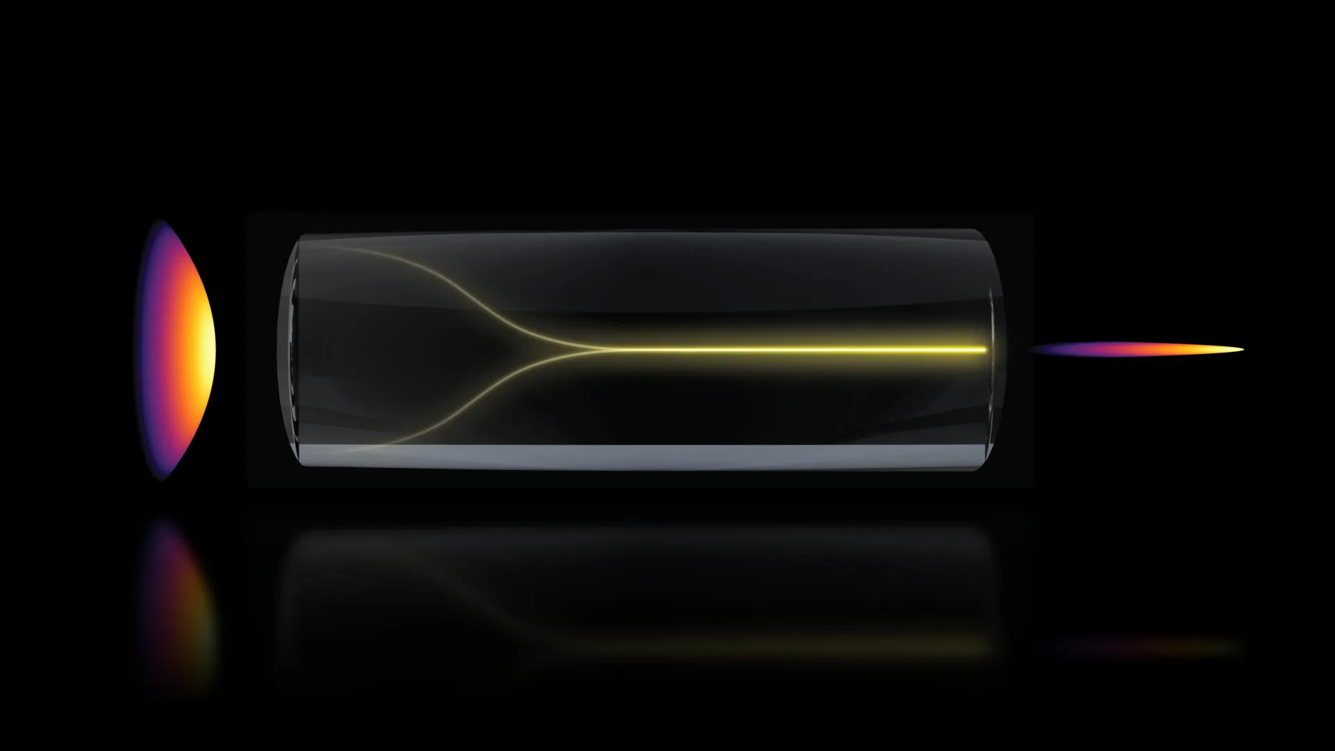
The landscape of optical physics and medical diagnostics has been fundamentally altered by a recent discovery at the Massachusetts Institute of Technology, where researchers have identified a phenomenon that allows laser light to self-organize into a highly focused "pencil beam." This unexpected effect, which occurs under specific high-power conditions within optical fibers, offers a transformative pathway for imaging living tissue with unprecedented speed and clarity. By harnessing this self-forming beam, an interdisciplinary team from MIT and Harvard University has successfully produced 3D images of the human blood-brain barrier at speeds 25 times faster than current industry standards, all while maintaining the high resolution necessary for cellular-level analysis. This breakthrough not only challenges long-held assumptions about the behavior of high-power lasers but also provides a critical tool for the pharmaceutical industry to track how drugs for neurodegenerative diseases, such as Alzheimer’s and ALS, interact with the brain in real time.
The Paradigm Shift in Optical Physics
For decades, the consensus among optical physicists was that increasing the power of a laser traveling through a multimode optical fiber would inevitably lead to chaos. Multimode fibers are designed to carry high levels of energy, but their internal structure is inherently disordered. As light travels through these fibers, imperfections in the glass cause the signal to scatter, resulting in a blurred and unstable output. Conventional wisdom suggested that "cranking up the power" would only exacerbate this disorder, making the resulting light useless for high-precision imaging without the use of expensive and complex beam-shaping hardware.
However, the MIT research team, led by Sixian You, an assistant professor in the Department of Electrical Engineering and Computer Science (EECS), discovered that the opposite occurs at a specific threshold. Instead of devolving into a chaotic mess, the light signal undergoes a process of self-organization. This transition results in a "pencil beam"—a narrow, stable, and extremely sharp column of light that can penetrate biological tissue with high precision. This discovery was the result of a rigorous experimental process that embraced the unexpected, proving that nonlinearity within the fiber can actually be used to counter intrinsic disorder rather than contribute to it.
Chronology of the Discovery
The discovery began in the MIT Research Laboratory for Electronics (RLE), where graduate student Honghao Cao was testing the limits of a "fiber shaper." This device was originally designed to provide fine-tuned control over laser light as it traversed a multimode fiber. During a routine series of tests, Cao began incrementally increasing the laser’s power to observe how the fiber handled high-energy loads.
The team anticipated that the light would begin to degrade as the power levels approached the fiber’s damage threshold. Instead, at a critical juncture, the scattered light suddenly collapsed into a single, concentrated point. This transition was instantaneous and stable. Recognizing the potential of this phenomenon, the team spent months identifying the exact variables required to replicate the effect. They found that two conditions were non-negotiable: a perfectly aligned, zero-degree entry angle for the laser and a power level high enough to trigger a direct interaction between the light and the glass material of the fiber. This interaction, known in physics as a nonlinear effect, allowed the light to essentially "sculpt" its own path, overcoming the physical imperfections of the fiber.
Technical Mechanics: Overcoming the Resolution-Depth Tradeoff
In traditional optical imaging, researchers are constantly forced to navigate a "tradeoff" between resolution and depth of focus. To see deep into a piece of tissue, one typically has to sacrifice the ability to see fine details; conversely, high-resolution imaging is usually limited to very thin slices of a sample. The self-organized pencil beam breaks this paradigm. Because the beam remains tightly focused over a long distance without the "sidelobes" (blurred halos) that plague conventional laser beams, it can maintain cellular-level resolution even as it probes deeper into 3D structures.
The MIT team utilized this beam to implement a label-free imaging technique. Most current bioimaging requires the use of fluorescent tags—chemical markers attached to cells or drugs to make them visible under a microscope. While effective, these tags can alter the behavior of the biological molecules being studied, leading to data that may not perfectly reflect natural processes. The new MIT method allows for the visualization of proteins and drug compounds in their native state, providing a more accurate representation of biological interactions.
Imaging the Blood-Brain Barrier: A New Frontier for Drug Development
The most immediate and impactful application of this technology was demonstrated in the imaging of the human blood-brain barrier (BBB). The BBB is a highly selective semipermeable border of endothelial cells that prevents solutes in the circulating blood from non-selectively crossing into the extracellular fluid of the central nervous system. While this barrier protects the brain from toxins and pathogens, it also serves as a massive hurdle for modern medicine, blocking more than 95% of potential drug compounds from reaching brain tissue.
To study the BBB, scientists often use engineered tissue models. However, tracking the movement of drugs through these models in 3D has historically been a slow and laborious process. Traditional methods involve taking multiple 2D scans and stitching them together, a process that is often too slow to capture the dynamic, real-time absorption of drugs by individual cells.
Using the self-organized pencil beam, the MIT team was able to:
- Accelerate Imaging Speed: The system generated 3D images roughly 25 times faster than the current gold-standard approaches.
- Track Real-Time Absorption: For the first time, researchers could watch individual cells internalize proteins and drug-like compounds as it happened.
- Identify Cell-Specific Rates: The clarity of the images allowed the team to distinguish between different cell types within the barrier and measure the specific rate at which each type absorbed the target molecules.
Roger Kamm, the Cecil and Ida Green Distinguished Professor of Biological and Mechanical Engineering at MIT, emphasized that this capability is a "game-changer" for the pharmaceutical industry. By using human-based tissue models instead of animal models—which often fail to accurately predict human responses—and removing the need for fluorescent tags, drug developers can get a much clearer picture of whether a treatment for Alzheimer’s or ALS is actually crossing the BBB and reaching the intended neurons.
Supporting Data and Comparative Analysis
The data published in Nature Methods highlights the efficiency of the new system. In comparative tests against standard multiphoton microscopy, the pencil beam approach showed a significant reduction in optical artifacts. While conventional beams often suffer from a loss of focus as they move away from the central axis, the self-organized beam remained stable.
Furthermore, the speed increase is not merely a marginal improvement but a step-function change. In the context of high-throughput drug screening, where thousands of compounds must be tested, a 25-fold increase in imaging speed could reduce the time required for certain stages of preclinical trials from months to weeks. This has profound implications for the cost of drug development, which currently averages over $2 billion per successful new drug brought to market.
Broader Implications and Future Research
The implications of this discovery extend far beyond the blood-brain barrier. Sarah Spitz, a postdoc on the project, noted that the method enables time-resolved tracking of diverse compounds across a wide array of engineered tissue models. This could include imaging the gut-brain axis, studying the way cancer cells metastasize through the vascular system, or observing the growth of lab-grown "organoids."
The simplicity of the setup is also a significant factor in its potential for widespread adoption. Because the effect can be achieved using standard optical components and does not require complex domain expertise in beam-shaping engineering, it is accessible to a broad range of biological laboratories.
Moving forward, the MIT team plans to delve deeper into the fundamental physics of the self-organization process. They aim to understand the exact mechanisms that allow the glass-light interaction to suppress disorder so effectively. Additionally, they are looking to adapt the technology for imaging live neurons in the brain, which could provide new insights into how neural circuits function and how they break down in the presence of disease.
Collaborative Effort and Funding
The success of this project was the result of a massive collaborative effort involving experts from MIT’s Department of Electrical Engineering and Computer Science, the Research Laboratory for Electronics, the Department of Biological Engineering, and Harvard University’s Beth Israel Deaconess Medical Center. Lead author Honghao Cao was joined by a diverse team including Li-Yu Yu, Kunzan Liu, Sarah Spitz, Francesca Michela Pramotton, Federico Presutti, Zhengyu Zhang, and Subhash Kulkarni.
The research was supported by a variety of prestigious institutions and funds, including the National Science Foundation (NSF), the Silicon Valley Community Foundation, the Diacomp Foundation, and the Harvard Digestive Disease Core. Additional support came from MIT startup funds, a MathWorks Fellowship, and the Claude E. Shannon Award.
As this technology moves from the lab toward practical application, it stands as a testament to the power of following unexpected evidence. By embracing the "chaos" of high-power lasers, the MIT team has found a way to let light organize itself, opening a new window into the microscopic world of the human body and offering hope for more effective treatments for some of the world’s most devastating diseases.
