
 By Suherman Candra Maholtra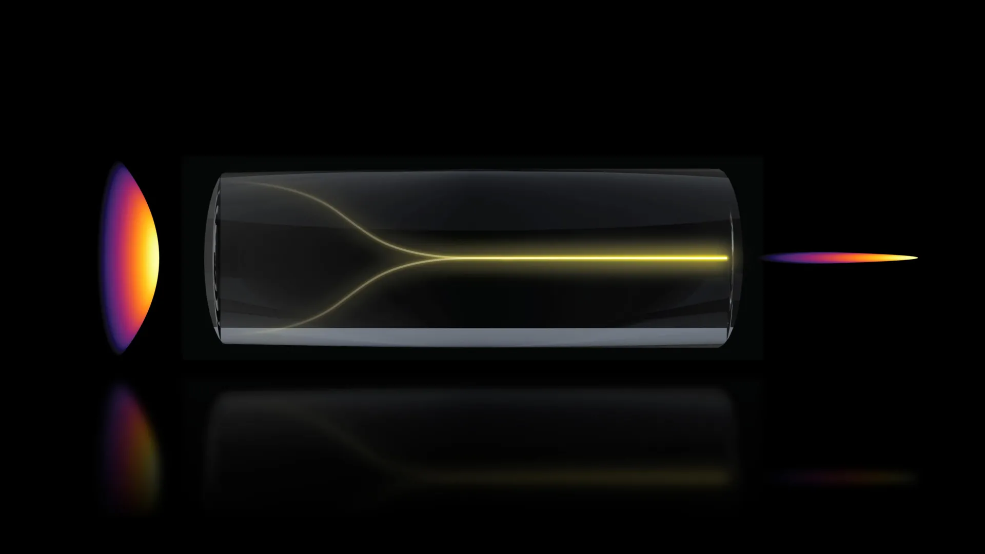
In a significant departure from long-held principles in optical physics, researchers at the Massachusetts Institute of Technology (MIT) have uncovered a phenomenon where high-power laser light, typically prone to chaotic scattering, spontaneously reorganizes into a highly focused "pencil beam." This discovery, detailed in a recent publication in Nature Methods, provides a transformative approach to biological imaging, allowing scientists to visualize complex structures like the human blood-brain barrier in three dimensions at speeds 25 times faster than current gold-standard technologies. By leveraging the intrinsic properties of light and glass, the team has bypassed traditional engineering hurdles, offering a new window into how life-saving drugs interact with the most protected regions of the human body.
A Paradigm Shift in Nonlinear Optics
For decades, the consensus among optical engineers was that increasing the power of a laser traveling through a multimode optical fiber would inevitably lead to increased disorder. Multimode fibers are designed to carry high levels of energy, but they are plagued by internal imperfections and "mode dispersion," which causes light to scatter into a disorganized pattern. Conventional wisdom suggested that to maintain a clean, usable beam at high power, researchers would need to employ complex, custom-made beam-shaping components or advanced computational corrections.
However, the MIT team, led by Sixian You, an assistant professor in the Department of Electrical Engineering and Computer Science (EECS), found that the light itself could provide the solution. By pushing the boundaries of laser power within a precisely aligned system, they observed the light overcoming its own disorder. Instead of diffusing into a blurred signal, the photons interacted with the molecular structure of the fiber’s glass to form a stable, ultra-sharp beam. This "self-organization" represents a milestone in nonlinear optics, proving that high-energy environments can produce order rather than chaos.
The Discovery: From Laboratory Accident to Scientific Breakthrough
The path to this discovery began with the development of a "fiber shaper," a device intended to provide granular control over laser light as it traverses multimode fibers. Lead author Honghao Cao, a graduate student in EECS, was testing the physical limits of these fibers by incrementally increasing the laser’s power. The expectation was that the fiber would eventually reach a breaking point where the image quality would degrade entirely or the fiber itself would sustain thermal damage.
As the power levels approached the damage threshold, the research team noticed a sudden and dramatic shift. The scattered light began to collapse inward, concentrating into a single, intense point of focus. This was not a fluke of the equipment but a fundamental physical response. The researchers identified that when the laser enters the fiber at a perfect zero-degree angle and reaches a "critical power," the nonlinearity of the glass begins to counter the intrinsic disorder of the fiber.
"Disorder is intrinsic to these fibers," Professor You explained. "The light engineering you typically need to do to overcome that disorder, especially at high power, is a longstanding hassle. But with this self-organization, you can get a stable, ultrafast pencil beam without the need for custom beam-shaping components."
Technical Specifications and the "Pencil Beam" Advantage
The resulting "pencil beam" is characterized by its high resolution and an unusually large depth of focus. In traditional microscopy, there is a fundamental trade-off: to see a cell in high detail, the microscope must focus on a very thin "slice" of the tissue. To see deeper, one usually has to sacrifice clarity. The MIT-developed beam maintains its tightness over a longer distance, allowing for deep-tissue penetration without the blurring common in other high-speed imaging methods.
Furthermore, the team’s approach eliminates "sidelobes"—the blurred halos that often surround laser beams in high-power applications. By producing a "clean" beam, the researchers can capture images with significantly fewer artifacts. This clarity is essential when attempting to distinguish between individual cells and the microscopic gaps between them in dense biological environments.
Visualizing the Blood-Brain Barrier in Real-Time
To demonstrate the practical utility of this discovery, the researchers applied the technique to one of the most challenging structures in human biology: the blood-brain barrier (BBB). The BBB is a highly selective semipermeable border of endothelial cells that prevents solutes in the circulating blood from non-selectively crossing into the central nervous system. While this barrier protects the brain from toxins and pathogens, it also serves as a massive obstacle for pharmaceutical treatments for neurodegenerative diseases.
Current methods for studying the BBB often rely on 2D imaging, which requires scanning multiple layers of tissue and digitally stitching them together to create a 3D model. This process is time-consuming and often fails to capture the dynamic movement of molecules in real-time.
Using the self-organized pencil beam, the MIT team was able to:
- Accelerate Imaging Speeds: The system produced 3D images roughly 25 times faster than the current gold-standard approach.
- Eliminate Fluorescent Tags: Traditional imaging often requires "tagging" drugs or cells with glowing chemicals to make them visible. The new MIT method can visualize these structures without tags, ensuring that the biological behavior being observed is natural and unaltered by the imaging process.
- Track Drug Absorption: For the first time, researchers could watch in real-time as individual cells within a human-based BBB model absorbed proteins and drugs.
Implications for Alzheimer’s and ALS Research
The ability to watch drugs cross the blood-brain barrier in real-time has profound implications for the pharmaceutical industry. Currently, many drugs that show promise in animal models for treating Alzheimer’s disease or Amyotrophic Lateral Sclerosis (ALS) fail in human clinical trials because they cannot effectively penetrate the human BBB.
Roger Kamm, the Cecil and Ida Green Distinguished Professor of Biological and Mechanical Engineering at MIT and a co-author of the study, noted that the industry is shifting toward human-based engineered tissue models to screen drugs more accurately. "That this new method doesn’t require the cells to have a fluorescent tag is a game-changer," Kamm said. "For the first time, we can now visualize the time-dependent entry of drugs into the brain and even identify the rate at which specific cell types internalize the drug."
This level of detail allows scientists to determine not just if a drug is reaching the brain, but how it is being processed once it arrives. This data is crucial for refining dosages and improving the molecular design of treatments to ensure they hit their intended targets.
A Collaborative Effort Across Disciplines
The research was a multi-disciplinary effort, reflecting the intersection of electrical engineering, biological engineering, and clinical medicine. The paper, published in Nature Methods, included contributions from EECS graduate students Li-Yu Yu and Kunzan Liu; postdocs Sarah Spitz, Francesca Michela Pramotton, and Federico Presutti; and Zhengyu Zhang.
The project also involved Subhash Kulkarni, an assistant professor at Harvard University and the Beth Israel Deaconess Medical Center, highlighting the collaborative nature of Boston’s scientific community. The diverse expertise of the team allowed them to move rapidly from the discovery of a physical phenomenon to its application in a complex biological setting.
Sarah Spitz emphasized that the utility of the pencil beam extends far beyond the brain. "This approach is not limited to the blood-brain barrier but enables time-resolved tracking of diverse compounds and molecular targets across engineered tissue models," she said. This suggests future applications in oncology, where researchers could track the penetration of chemotherapy into solid tumors, or in cardiology, to study the absorption of nutrients in heart tissue.
Future Directions and Commercial Potential
While the current results are groundbreaking, the MIT team views this as the beginning of a new era in optical imaging. The next phase of research will focus on two primary objectives: deepening the understanding of the underlying physics and expanding the clinical applications.
Physically, the researchers want to map the exact mechanisms that allow the glass material of the fiber to interact with high-power light to create order. Understanding these "nonlinear interactions" could allow engineers to design even more efficient fibers that require less power to trigger the self-organizing effect.
Clinically, the team aims to use the method to image neurons in action, potentially providing new insights into how the brain’s electrical signals change in response to disease. There is also significant interest in commercializing the technology. Because the method uses standard optical setups and does not require "domain expertise" in complex beam-shaping, it could be easily integrated into existing laboratory workflows across the globe.
Funding and Institutional Support
The research received support from a wide array of prestigious organizations, including the National Science Foundation (NSF) and the Silicon Valley Community Foundation. Additional funding was provided by MIT startup funds, the Diacomp Foundation, the Harvard Digestive Disease Core, a MathWorks Fellowship, and the Claude E. Shannon Award.
As the pharmaceutical industry continues to seek more efficient ways to bridge the gap between laboratory research and human cures, the MIT "pencil beam" stands as a testament to the power of following the evidence—even when it contradicts long-held scientific beliefs. By embracing the "chaos" of high-power lasers, these researchers have illuminated a faster, clearer path toward understanding the most complex systems of the human body.
