
 By Suherman Candra Maholtra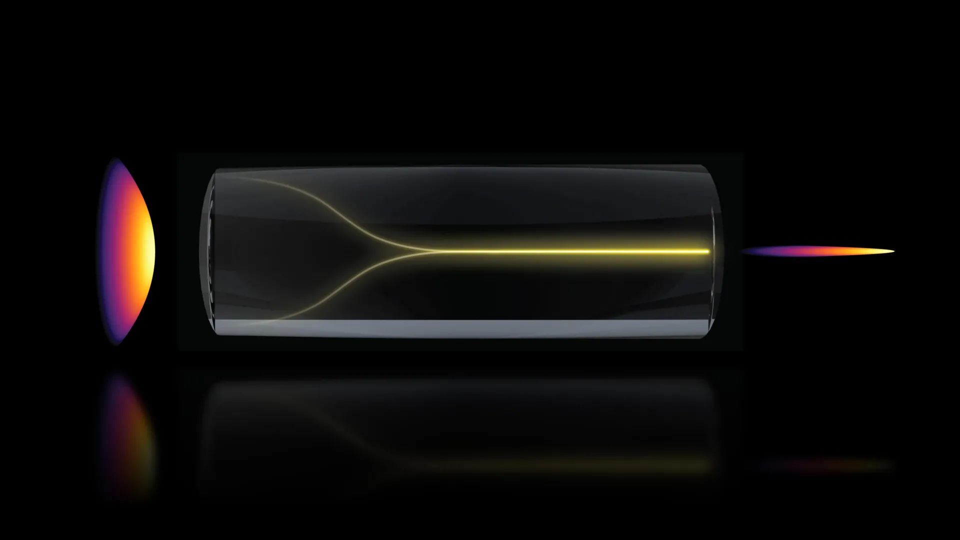
In a discovery that challenges long-standing conventions in optical physics, a research team at the Massachusetts Institute of Technology (MIT) has identified a phenomenon where chaotic laser signals spontaneously reorganize into a highly focused "pencil beam." This self-organizing light effect, previously thought impossible under high-power conditions in multimode fibers, provides a transformative pathway for biological imaging. By leveraging this stable, narrow beam, researchers have successfully produced three-dimensional images of the human blood-brain barrier at speeds approximately 25 times faster than current gold-standard technologies. The breakthrough, published in the journal Nature Methods, offers a label-free method to observe cellular processes in real-time, potentially revolutionizing how the pharmaceutical industry evaluates treatments for neurodegenerative diseases such as Alzheimer’s and Amyotrophic Lateral Sclerosis (ALS).
The Mechanics of Spontaneous Optical Reorganization
The discovery centers on the behavior of light within multimode optical fibers. Traditionally, these fibers are utilized for their ability to carry high levels of power, but they are notorious for "modal dispersion"—a phenomenon where different rays of light travel at different speeds and paths, resulting in a scrambled, speckled output. For decades, the prevailing wisdom among physicists was that increasing the power input into such a system would only exacerbate this chaos, leading to a breakdown of signal integrity.
However, the MIT team, led by Assistant Professor Sixian You and graduate student Honghao Cao, found that under specific "Goldilocks" conditions, the system undergoes a phase transition. When a laser is injected into a fiber at a precise zero-degree angle and the power is increased toward the fiber’s damage threshold, the intrinsic nonlinearity of the glass begins to interact with the light. Instead of shattering into a disordered pattern, the light collapses into a singular, ultra-sharp "pencil beam."
This self-organization occurs because the nonlinear effects of the medium act as a corrective force, countering the natural disorder of the fiber. This creates a stable, high-intensity beam that maintains a large depth of focus without the need for the complex and expensive spatial light modulators or custom beam-shaping hardware typically required in advanced microscopy.
A Chronology of the Discovery
The path to this discovery was rooted in a series of experiments designed to test the physical limits of a "fiber shaper," a device the team had previously engineered to control light propagation. During a routine testing phase, lead author Honghao Cao began incrementally increasing the laser power to determine the maximum capacity of the multimode fiber.
In a typical setting, as the power climbs, the light output becomes increasingly "noisy" due to microscopic imperfections in the fiber’s silica core. However, as Cao approached a critical power threshold—just below the point where the fiber would physically melt or fracture—the speckled pattern on the laboratory monitors suddenly condensed into a brilliant, concentrated point of light.
Recognizing that they had stumbled upon a fundamental physical exception, the team spent months replicating the effect. They identified two non-negotiable parameters for the phenomenon: near-perfect axial alignment of the input laser and a high-intensity power state that triggers the glass’s nonlinear refractive properties. By "embracing the uncertainty" of the data, as Professor You described it, the team moved from an accidental observation to a controlled, reproducible imaging technique.
Technical Specifications and Comparative Performance
The primary advantage of the "pencil beam" lies in its unique combination of high resolution and extended depth of focus. In traditional optical imaging, there is an inverse relationship between these two factors: a beam focused sharply on a single point (high resolution) typically has a very shallow depth of field, meaning only a thin slice of tissue can be imaged at once. To create a 3D image, a microscope must mechanically scan through hundreds of these thin slices, a process that is both time-consuming and prone to motion artifacts.
The MIT-developed pencil beam bypasses this trade-off. Because the beam remains narrow over a long distance, it can capture data from multiple depths simultaneously or with significantly fewer scans.
Key Performance Metrics:
- Imaging Speed: The system achieves 3D visualization 25 times faster than traditional point-scanning methods like two-photon microscopy or optical coherence tomography (OCT) in similar biological contexts.
- Beam Quality: Unlike conventional "Bessel beams" or other engineered light shapes, which often produce "sidelobes" (halos of light that blur the image), the self-organized beam is "clean," resulting in higher contrast and fewer artifacts.
- Label-Free Imaging: The technique does not require the use of fluorescent dyes or chemical tags. This is a critical development for medical research, as chemical tags can sometimes alter the behavior of the cells being studied.
Real-Time Visualization of the Blood-Brain Barrier
To demonstrate the practical utility of this physics breakthrough, the researchers applied the technique to one of the most challenging structures in human biology: the blood-brain barrier (BBB). The BBB is a highly selective semipermeable border of endothelial cells that prevents solutes in the circulating blood from non-selectively crossing into the extracellular fluid of the central nervous system.
While the BBB protects the brain from toxins, it also serves as a massive hurdle for drug delivery. Approximately 98% of small-molecule drugs and nearly 100% of large-molecule drugs (such as those for Alzheimer’s) fail to cross this barrier. Using the new imaging method, the MIT team was able to watch individual cells within an engineered human BBB model as they interacted with various proteins and drug-mimicking compounds.
For the first time, researchers could see the time-dependent entry of these substances into the brain tissue. They were able to identify the specific rates at which different cell types internalized compounds, providing a level of granular data that was previously inaccessible. This capability is particularly vital for the pharmaceutical industry, which currently relies heavily on animal models that often fail to replicate the complexities of the human blood-brain barrier.
Implications for Drug Development and Bioengineering
The ability to track drug absorption in real-time without damaging the tissue or using invasive tags has profound implications for clinical research. Professor Roger Kamm, a co-author of the study and a specialist in biological engineering, noted that the pharmaceutical industry is in desperate need of human-based models to screen for drugs. By providing a high-speed, high-resolution window into the BBB, this technology could significantly reduce the time and cost associated with drug development for neurological disorders.
Beyond the brain, the "pencil beam" technique is expected to be versatile. Sarah Spitz, a postdoc on the project, emphasized that the method is not limited to the BBB. It can be applied to any engineered tissue model to track molecular targets and diverse compounds across various organs-on-a-chip. This makes it a powerful tool for the broader field of biological engineering, including cancer research and regenerative medicine.
Future Research and Commercial Potential
The MIT team is now focused on two parallel tracks: deepening the theoretical understanding of the physics involved and practical miniaturization of the technology. While they have successfully identified the conditions required for the pencil beam to form, the exact molecular-level interactions between the high-power photons and the silica lattice of the fiber remain a subject of intense study.
On the application side, the researchers are looking to adapt the system for imaging neurons and other deep-tissue structures that are typically difficult to reach with light. Because the method uses standard multimode fibers and does not require complex beam-shaping components, it is inherently more cost-effective and easier to implement than many existing high-end microscopy systems. This "simplicity" suggests a high potential for commercialization and widespread adoption in both academic and industrial laboratories.
Acknowledgments and Institutional Support
The interdisciplinary effort involved researchers from MIT’s Department of Electrical Engineering and Computer Science (EECS), the Research Laboratory for Electronics (RLE), the Department of Biological Engineering, and Harvard University’s Beth Israel Deaconess Medical Center.
The research was supported by a diverse array of funding bodies, reflecting its cross-disciplinary impact. Contributors include the National Science Foundation (NSF), MIT startup funds, the Silicon Valley Community Foundation, the Diacomp Foundation, and the Harvard Digestive Disease Core. Individual researchers were also supported by the MathWorks Fellowship and the Claude E. Shannon Award.
As the paper appears in Nature Methods, it marks a significant milestone in the intersection of nonlinear optics and biomedical imaging, proving that sometimes, the most elegant solutions in science are found by pushing systems to the very edge of chaos.
