
 By Nana O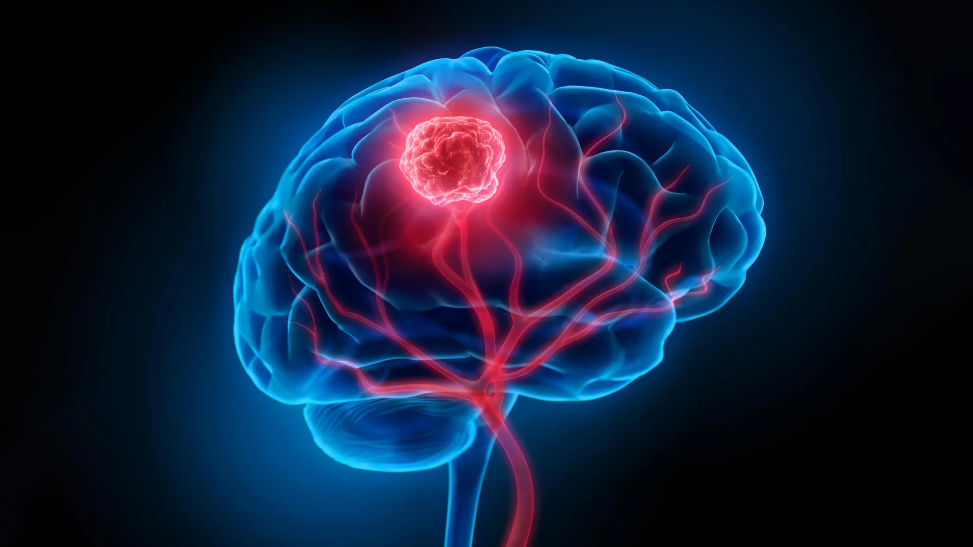
Glioblastoma, a notoriously aggressive form of brain cancer, has long presented a formidable challenge to medical science, with limited treatment options and grim prognoses. However, a groundbreaking development from a collaborative team of researchers at the University of Technology Sydney (UTS), Harvard University, and Henan University may herald a significant shift in how this devastating disease is tackled. Their innovative ‘double-punch’ nanozyme platform, detailed in the prestigious journal Science Translational Medicine, offers a dual-action approach designed to overcome two of the most significant obstacles in glioblastoma therapy: the infiltrative nature of cancer cells and the protective blood-brain barrier.
The inherent difficulty in treating glioblastoma stems from its insidious spread. Cancer cells permeate surrounding healthy brain tissue, making complete surgical excision a delicate and often incomplete process. Surgeons must navigate a complex landscape, meticulously removing cancerous tissue while preserving vital neurological functions. This microscopic infiltration, even after apparent gross tumor removal, is a primary driver of recurrence. Compounding this challenge is the blood-brain barrier (BBB), a highly selective physiological shield that protects the brain from circulating toxins but also severely restricts the delivery of therapeutic agents, including chemotherapy drugs and the effectiveness of radiotherapy. The confluence of these factors contributes to the stark reality that the five-year survival rate for glioblastoma patients remains tragically low, hovering around a mere 7 percent.
A Dual-Action Nanoparticle System: The ‘Double-Punch’ Approach
The newly developed nanozyme platform represents a paradigm shift in treatment strategy, employing a single, intelligent system of nanoparticles to perform two critical functions sequentially. Dr. Bingyang Shi, Chair Professor of Nanomedicine at UTS, who led the research, explained the ingenious design: "We’ve engineered a single material that does two jobs in sequence. It’s a precise guide for the surgeon during the operation, and then a targeted clean-up treatment afterward." This elegant solution aims to empower surgeons with enhanced visualization during procedures and subsequently provide a potent, localized therapeutic intervention to eliminate any residual cancerous cells.
At the heart of this innovative system lies an ultra-thin, two-dimensional sheet composed of individual atoms meticulously arranged. This fabrication process, adapted from advanced semiconductor manufacturing techniques, imbues the material with the remarkable ability to toggle between distinct functionalities. Crucially, both of these functions are activated by the same near-infrared (NIR) light, simplifying the therapeutic protocol.
Enhanced Surgical Precision: Illuminating Microscopic Tumors
The first critical role of the nanoparticle platform is as a highly sensitive imaging agent during surgery. "During surgery, it functions as a highly sensitive imaging agent," stated Professor Shi. Engineered with a fluorescent dye, the material emits a glow under NIR light, a wavelength invisible to the human eye. This allows surgeons to visualize individual tumor cell clusters that are as small as 44 micrometers – a resolution that surpasses the capabilities of current clinical imaging tools. This enhanced visibility is paramount for maximizing tumor resection while minimizing damage to healthy brain tissue.
Furthermore, the nanoparticles are equipped with a targeting molecule designed to actively cross the blood-brain barrier. This crucial feature ensures that the imaging agent accumulates specifically within glioma cells, effectively highlighting the cancerous margins of the tumor and distinguishing them from surrounding healthy brain matter. This precision guidance is expected to lead to more complete initial surgical removals, a critical factor in improving patient outcomes.
Targeted Post-Operative Therapy: Eliminating Residual Cancer Cells
Following the surgical removal of the visible tumor, the same nanoparticle platform is deployed within the surgical cavity. Upon reactivation with the same NIR light, the material transitions to its therapeutic role. The embedded platinum atoms play a key part in this phase. They catalyze a reaction that converts the tumor’s own hydrogen peroxide into oxygen. This process is vital, as glioblastoma tumors often create a hypoxic (low-oxygen) microenvironment, which typically shields cancer cells from treatment. By increasing oxygen levels, the nanoparticles counteract this protective mechanism.
Simultaneously, the NIR light triggers the generation of heat and reactive oxygen species. These localized effects are designed to destroy any microscopic cancer cells that may have escaped the surgeon’s notice and remained in the brain. This ‘clean-up’ phase is crucial for preventing tumor recurrence, a major cause of mortality in glioblastoma patients. The ability of a single material to perform both imaging and therapy in a sequential, targeted manner represents a significant advancement over current multimodal treatment strategies.
Pre-Clinical Success: Promising Results in Animal Models
The efficacy of this novel nanoparticle platform has been demonstrated in pre-clinical studies using mouse models of glioblastoma. The results are highly encouraging. In these models, the nanoparticle-assisted treatment significantly reduced tumor recurrence following surgery. Notably, all mice treated with the nanoparticle approach remained alive at the 60-day mark, a substantial improvement compared to the 42-day survival observed in control mice that underwent surgery alone.
Crucially, follow-up assessments revealed no detectable neurological or motor impairments associated with the nanoparticle treatment, suggesting a favorable safety profile in these animal models. This absence of adverse effects is a critical factor for any therapeutic intervention in the brain, where even subtle functional deficits can have a profound impact on a patient’s quality of life.
Background and Context: The Unrelenting Challenge of Glioblastoma
Glioblastoma (GBM) is the most common and aggressive primary malignant brain tumor in adults, accounting for approximately 15% of all primary brain tumors. The World Health Organization (WHO) classifies it as a Grade IV astrocytoma, signifying its rapid growth and invasive nature. The median survival for patients diagnosed with glioblastoma has historically been grim, often measured in months rather than years, despite aggressive treatment regimens that typically involve surgery, radiation therapy, and chemotherapy.
The complex biology of glioblastoma, characterized by its diffuse infiltration, genetic heterogeneity, and resistance to conventional therapies, has made it a particularly challenging disease to treat. Over the decades, numerous research efforts have aimed to improve surgical techniques, develop more effective chemotherapies, and explore novel approaches like immunotherapy and targeted therapies. However, the inherent limitations of the blood-brain barrier and the infiltrative nature of the tumor have consistently hampered progress.
The development of advanced imaging techniques and targeted drug delivery systems has been a key focus in glioblastoma research. Technologies that can accurately delineate tumor margins during surgery and deliver therapeutic agents directly to cancer cells while sparing healthy tissue are highly sought after. The UTS-led initiative directly addresses these critical needs by integrating both imaging and therapeutic capabilities within a single, sophisticated nanoparticle system.
Implications and Future Directions: A Step Towards Personalized Treatment
The implications of this research are substantial. If successfully translated to human clinical trials, this ‘double-punch’ nanozyme platform could revolutionize glioblastoma surgery and post-operative care. Surgeons could benefit from unprecedented visualization, leading to more complete tumor resections. The subsequent targeted therapy would then systematically eliminate any remaining microscopic cancer cells, significantly reducing the likelihood of recurrence, which is currently a major cause of patient mortality.
Professor Shi emphasized the importance of continued research and validation: "The results are very encouraging, but this is still early-stage research carried out in mouse models, not in people – and that distinction is important." The transition from animal models to human trials is a complex and lengthy process, involving rigorous safety and efficacy testing. The performance of the nanoparticles will need to be confirmed at the scale of a human brain, and potential immunogenic responses in human patients will need to be carefully evaluated.
However, the potential impact is clear. "If this continues to hold up through that process, the hope is that surgeons could one day see more of the tumor during an operation and treat more of what’s left behind afterward," Professor Shi stated. "It’s a meaningful step towards reducing recurrence, which remains one of the biggest challenges for people with glioblastoma."
The research team is optimistic about the future. The ability to precisely target and treat glioblastoma cells, both during surgery and in the post-operative period, could pave the way for more personalized and effective treatment strategies. This breakthrough represents a beacon of hope for patients and clinicians grappling with one of the most formidable cancers known to medicine, potentially ushering in an era where glioblastoma is no longer an insurmountable diagnostic and therapeutic hurdle. The continued collaboration between institutions like UTS, Harvard, and Henan universities underscores the global effort to combat this devastating disease through interdisciplinary innovation and scientific advancement.
