
 By Budi Oetomo Narendra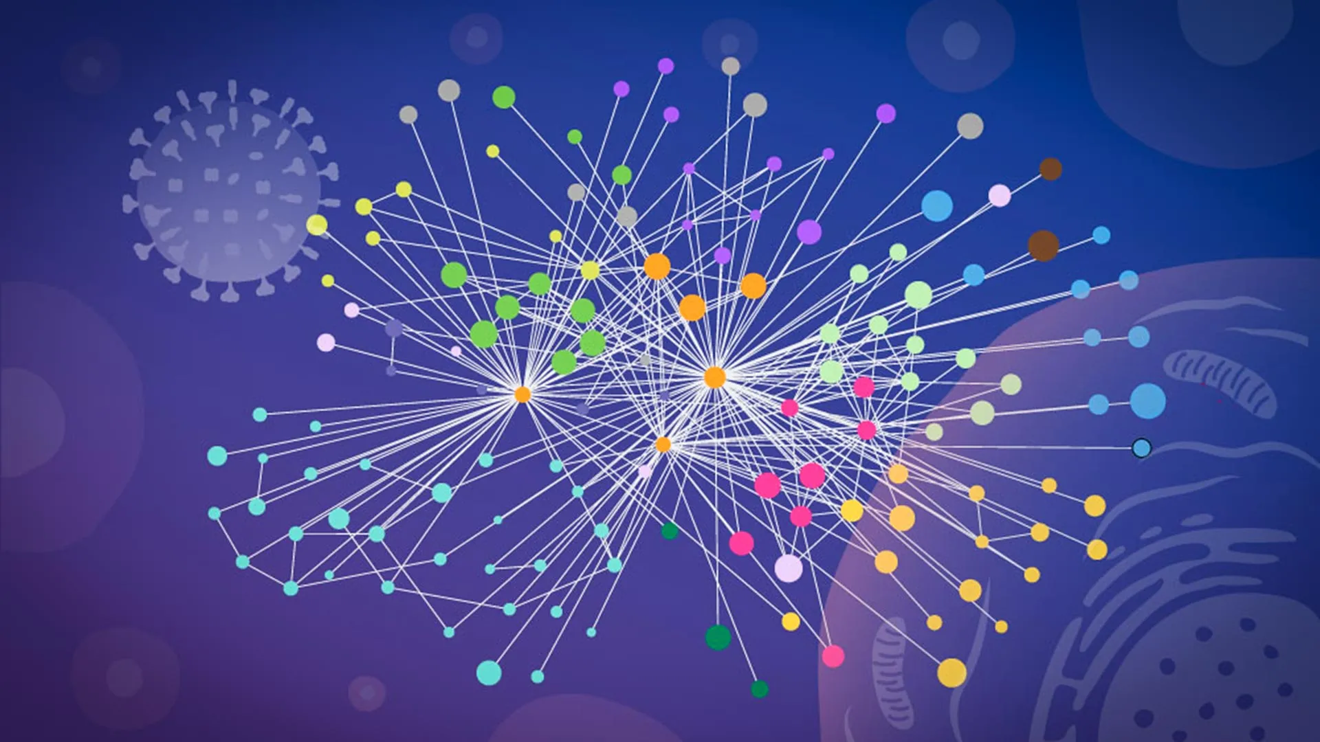
The Persistent Global Threat of Influenza A
Influenza A stands as a formidable and perennial threat to global public health. Seasonal influenza, primarily driven by types A and B, is responsible for an estimated 3 to 5 million cases of severe illness and between 290,000 and 650,000 respiratory deaths worldwide annually, according to the World Health Organization (WHO). Influenza A viruses are particularly concerning due to their capacity for rapid evolution, undergoing both antigenic drift (minor genetic changes) and antigenic shift (major genetic reassortment), which can lead to new viral strains capable of evading existing immunity and causing pandemics.
Historically, influenza A has been the architect of some of humanity’s most devastating pandemics. The most infamous example is the 1918 "Spanish Flu" pandemic, caused by an H1N1 influenza A virus, which infected an estimated one-third of the world’s population and claimed at least 50 million lives globally. More recently, the 2009 H1N1 "Swine Flu" pandemic, also an influenza A strain, demonstrated the continued potential for widespread disruption and mortality, even in the age of modern medicine and rapid vaccine development. Current concerns also revolve around highly pathogenic avian influenza (HPAI) strains like H5N1 and H7N9, which, while primarily affecting birds, have shown sporadic transmission to humans with high fatality rates, raising fears of their potential to adapt for efficient human-to-human transmission and trigger future pandemics. Understanding the precise mechanisms by which influenza A hijacks human cells is thus not merely an academic exercise but a critical endeavor in public health defense.
The Intricate Viral Takeover: A Molecular Battleground
Upon entering a human cell, the influenza A virus initiates a sophisticated takeover operation. It releases its segmented RNA genome, which contains the genetic blueprints for a small but potent arsenal of viral proteins. These proteins then fan out across the host cell, systematically subverting and redirecting its complex molecular machinery. The cell, once a functional unit of the human body, is transformed into a veritable factory dedicated solely to the production of new virus particles. This process, known as viral replication, is a tightly choreographed series of interactions between viral proteins and hundreds of host cellular proteins.
A clearer, more granular understanding of this intricate takeover process is essential for developing next-generation flu vaccines and more effective antiviral drugs. To achieve this, scientists need to precisely identify which viral proteins interact with which human proteins, pinpoint the exact cellular locations where these contacts occur, and elucidate the molecular mechanisms by which the virus leverages these interactions to support its own replication cycle. Traditional research methods have often fallen short in providing this level of detail, particularly regarding the dynamic and spatially restricted nature of these interactions within a living cell.
Overcoming Methodological Hurdles: The Need for In Situ Mapping
Tracking protein-protein interactions (PPIs) during an active viral infection presents immense experimental challenges. Many earlier studies relied on biochemical techniques that necessitated the physical disruption of cells—a process known as cell lysis—before protein contacts could be measured. While valuable, this approach carries significant limitations.
The act of breaking cells open fundamentally alters the cellular environment. Internal compartments, such as the nucleus, endoplasmic reticulum, or Golgi apparatus, are destroyed, leading to a homogenization of cellular contents. This can result in the formation of artificial protein contacts between molecules that were originally separated within the living cell, leading to false positives. Conversely, weak, transient, or highly localized interactions—which are often critical in dynamic biological processes like viral infection—can easily be disrupted or lost during cell lysis, leading to false negatives. Consequently, researchers have often struggled to definitively determine which connections truly existed and were functionally relevant during the course of a genuine infection. The scientific community recognized a pressing need for methodologies that could capture these interactions in situ, preserving the native cellular context and providing a more accurate snapshot of the viral takeover.
A Technological Leap: Specialized Cross-linking Mass Spectrometry
The breakthrough in addressing this challenge came through a pivotal collaboration. "This is when we learned that our collaborators, Boris Bogdanow and Fan Liu, at FMP Berlin had developed a specialized version of cross-linking mass spectrometry (XL-MS), a long-established technique for mapping protein contacts, tailored specifically to virus-infected cells," stated Jan Kosinski, Group Leader at EMBL Hamburg and Centre for Structural Systems Biology (CSSB). This specialized method proved to be the crucial innovation the team required.
Cross-linking mass spectrometry is a powerful biochemical technique that uses chemical reagents to create covalent bonds (cross-links) between proteins that are in close proximity within a sample. These cross-links essentially "freeze" the interactions in place, allowing researchers to later identify the linked proteins and, critically, the specific amino acid residues involved in the interaction. The unique adaptation developed by Bogdanow and Liu for virus-infected cells dramatically enhanced the utility of XL-MS. It allowed them to capture interactions that occur only briefly or within particular, often dynamic, regions of an infected cell, providing an unprecedented level of spatial and temporal resolution.
"XL-MS allows us to capture protein-protein interactions directly in infected intact cells, while also providing structural information about how these interactions are happening," explained Bogdanow, who is now a Junior Research Group Leader at the Institute of Virology, Charité — Universitätsmedizin Berlin. "This gives us insight into the interface between the virus and the human cell and may, through structural modeling, help identify actionable targets for future pharmaceutical interventions." This capability to directly observe and structurally characterize interactions within the native cellular environment represented a significant leap forward in understanding viral pathogenesis.
The Power of AI: Integrating Experimental Data with AlphaFold
To further enrich their findings and move beyond simply identifying interacting proteins, the researchers paired their high-resolution XL-MS results with advanced computational structural modeling. This synergistic approach allowed them not only to pinpoint which viral and human proteins interact but also to estimate with remarkable precision how those proteins are spatially positioned and fit together when they connect.
To construct these detailed structural models, the team leveraged a modified version of AlphaFold, the revolutionary protein structure prediction algorithm developed by DeepMind. AlphaFold has garnered widespread acclaim for its ability to accurately predict the 3D structures of proteins solely from their amino acid sequences, a feat that was once considered a grand challenge in biology. While AlphaFold itself is a powerful predictive tool, its true potential is maximized when integrated with experimental data, especially for complex systems like virus-host interactions.
"The key advantage of the modified AlphaFold approach is that it allowed us to feed our experimental cross-linking data directly into the structural modeling," explained Kosinski. "This tells the model which parts of the viral and host proteins are close to each other inside infected cells. This was especially useful for virus-host complexes, which are often difficult to predict reliably." By incorporating empirical cross-link constraints, the modified AlphaFold algorithm was guided by real-world interaction data, significantly enhancing the accuracy and biological relevance of the predicted complex structures. This integration of cutting-edge experimental techniques with state-of-the-art artificial intelligence represents a new paradigm in structural biology.
Two Key Strategies of Influenza A Hijacking
The findings, recently published in the prestigious journal Nature Microbiology, illuminated two particularly notable and previously uncharacterized strategies that influenza A appears to employ when asserting control over a human cell. These discoveries provide crucial insights into the virus’s sophisticated manipulative tactics.
1. Hemagglutinin Processing and Host Protein Exploitation:
The first strategy centers on hemagglutinin (HA), a crucial glycoprotein found on the surface of the influenza virion. Hemagglutinin is indispensable for the initial stages of infection, acting as the primary molecule responsible for binding to sialic acid receptors on the host cell surface and facilitating viral entry through endocytosis. Following entry, the virus’s newly synthesized hemagglutinin proteins must be correctly folded, modified, and transported to the cell surface to form new viral particles.
The researchers meticulously tracked hemagglutinin’s journey through the host cell’s intricate internal transport and processing network, which includes the endoplasmic reticulum (ER) and Golgi apparatus. These cellular compartments are responsible for the synthesis, folding, modification (e.g., glycosylation), and quality control of proteins destined for secretion or insertion into membranes. The detailed analysis revealed that several human proteins within this network actively assisted in the correct folding and modification of hemagglutinin during infection. Intriguingly, some of these host proteins previously had poorly understood or ambiguous functions within the cell. This finding suggests that influenza A not only co-opts the host cell’s protein synthesis machinery but also its quality control and maturation systems, essentially turning the cell’s own resources against itself to ensure the production of functional viral components. Understanding which specific host factors are involved in HA maturation could present novel targets for antiviral drugs that disrupt this critical step in the viral life cycle.
2. Dissolution of Paraspeckles and Resource Mobilization:
The second, perhaps even more surprising, discovery involved paraspeckles. These are small, dynamic, droplet-like sub-compartments located within the cell nucleus, composed primarily of RNA and RNA-binding proteins. Paraspeckles are known to play roles in regulating gene expression, sequestering specific RNAs, and contributing to cellular stress responses and antiviral defense mechanisms.
The research team observed a consistent and dramatic phenomenon: influenza A infection caused these paraspeckle structures to dissolve. This dissolution was not an isolated event but occurred consistently across every cell line and every flu strain tested. When paraspeckles broke apart, they released their sequestered cargo, primarily various RNA-binding proteins that had been held within them. The critical implication here is that the virus may then commandeer these liberated RNA-binding proteins, utilizing them to support its own replication and transcription processes, which heavily rely on host RNA machinery.
"What surprised us most was the paraspeckles," said Iuliia Kotova, former predoctoral fellow at the Kosinski Group at EMBL Hamburg, currently at ETH Zurich and first author of the publication. "Watching these tiny organelles in the nucleus dissolve, consistently across every cell line and every flu strain we tested, told us this isn’t a side effect of infection — it might be a strategy." This observation strongly suggests a deliberate viral mechanism rather than a collateral damage effect.
The disruption of paraspeckles may provide influenza with more than one strategic advantage. "There may also be a second benefit for the virus: some evidence suggests paraspeckles contribute to cellular stress responses and antiviral gene regulation, so disrupting them could also weaken parts of the cell’s defense response," added Kosinski. By dismantling these nuclear defense outposts, influenza A could simultaneously acquire essential host factors for its own replication and suppress the host cell’s innate antiviral immunity, creating a more permissive environment for its proliferation. This dual benefit highlights the evolutionary sophistication of the virus.
A Collaborative Symphony Across Leading Institutions
This comprehensive and technically demanding project was a testament to the power of inter-institutional collaboration, drawing on shared technology and diverse expertise from three leading European research institutions. The cross-linking mass spectrometry work, a cornerstone of the study, was meticulously carried out at Charité — Universitätsmedizin Berlin, under the guidance of Boris Bogdanow. Simultaneously, critical glycoproteomics analyses, essential for understanding protein modifications like glycosylation, were expertly completed at the EMBL Proteomics Core Facility in Heidelberg.
The sophisticated AlphaFold modeling, which transformed raw interaction data into detailed structural insights, was performed on the high-performance EMBL Compute Cluster, leveraging its vast computational resources. Complementary microscopy imaging, providing visual confirmation and context for the molecular events, took place at the Centre for Structural Systems Biology (CSSB)’s state-of-the-art Advanced Light and Fluorescence Microscopy (ALFM) Facility. This multidisciplinary orchestration of specialized techniques and facilities underscores the collaborative nature of modern biological discovery, where no single lab or institution possesses all the necessary tools and expertise to tackle such complex biological questions.
Broader Implications and Future Directions for Pandemic Preparedness
The findings of this study extend far beyond influenza A, demonstrating a powerful new paradigm for understanding viral pathogenesis. By meticulously studying molecular contacts inside intact infected cells, the researchers have revealed not only where but also how a virus commandeers human cellular machinery. This type of "mapping in context" approach holds immense promise for explaining the operational strategies of a wide array of other viruses.
"While the exact host factors and mechanisms often differ from virus to virus, we think our overall approach — combining in-cell cross-linking, structural modeling, and targeted cell-biology follow-up to map native virus-host interactions at specific stages of infection — remains broadly applicable," Kosinski affirmed. This methodology could be adapted to investigate other significant human pathogens, including other respiratory viruses like SARS-CoV-2, HIV, Ebola, or Zika virus, each with their unique modes of host interaction.
Although the current study focused on a laboratory-adapted strain of influenza, the researchers are confident that the same rigorous strategy can eventually be deployed to investigate viruses with far greater pandemic potential. Bogdanow reiterated this potential: "Although this study has focused on a lab-adapted strain, this study lays the groundwork to apply the methodology to viruses of potential pandemic relevance, such as H5N1, and for uncovering the interaction networks that support their multiplication in human cells." Applying this advanced mapping technique to emerging viral threats, such as novel avian influenza strains or coronaviruses, could rapidly accelerate our understanding of their infectivity, pathogenicity, and species jump capabilities, providing crucial insights for proactive pandemic preparedness and the development of targeted countermeasures.
In conclusion, this landmark study not only provides an unprecedented, high-resolution view of influenza A’s intricate dance with human cells but also establishes a robust methodological framework for dissecting the molecular strategies of other viruses. By integrating cutting-edge experimental techniques with advanced computational modeling, researchers are now better equipped to uncover the vulnerabilities of viral pathogens, paving the way for a new generation of vaccines and antiviral drugs that could significantly reduce the global burden of infectious diseases.
