
 By Suherman Candra Maholtra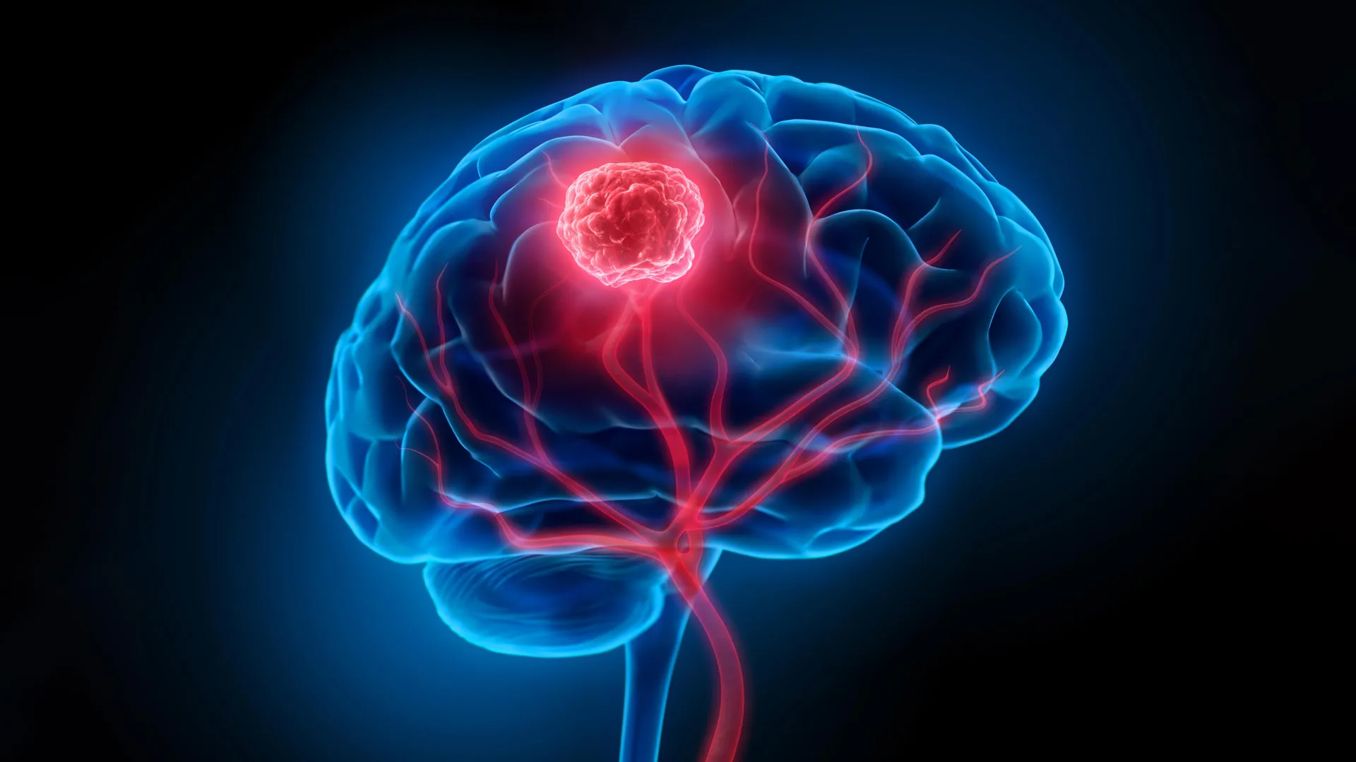
In a landmark discovery that challenges the traditional understanding of neuro-oncology, a collaborative research team in South Korea has identified the cellular origin of isocitrate dehydrogenase (IDH)-mutant glioma, the most prevalent malignant brain tumor among adults under the age of 50. The study, published on January 8th in the prestigious journal Science, reveals that these tumors do not emerge as sudden, localized masses but rather originate from Glial Progenitor Cells (GPCs) that quietly infiltrate the normal-looking cerebral cortex years before a tumor becomes visible on medical imaging. This finding explains the high recurrence rates of the disease and opens a new frontier for early diagnostic techniques and preventative therapies.
The research was led by Professor Jeong Ho Lee of the Korea Advanced Institute of Science and Technology (KAIST) Graduate School of Medical Science and Engineering, alongside Professor Seok-Gu Kang of Yonsei University Severance Hospital. By tracing the genetic lineage of the cancer back to its earliest mutations in otherwise healthy brain tissue, the team has provided a roadmap for moving beyond the "debulking" surgery model—which focuses on removing the visible tumor—toward a paradigm of early molecular intervention.
The Challenge of IDH-Mutant Glioma
Gliomas are tumors that arise from the glial cells, which support and protect neurons in the brain. Among these, IDH-mutant gliomas represent a specific subtype defined by a mutation in the IDH1 or IDH2 genes. While these tumors generally grow more slowly than the highly aggressive IDH-wildtype glioblastomas, they are notoriously difficult to cure. Patients often undergo successful surgery to remove the visible mass, only to face a recurrence years later, often in a more aggressive form.
Until now, the medical community lacked a clear understanding of where these tumors began and how they behaved in their "latent" phase. Standard clinical practice relies on Magnetic Resonance Imaging (MRI) to identify tumor boundaries. However, the South Korean team’s research suggests that by the time a mass is visible on an MRI, the mutation-carrying cells have already established a wide-reaching presence throughout the brain’s architecture.
Tracking the Invisible: The Role of Glial Progenitor Cells
The breakthrough centered on the identification of Glial Progenitor Cells (GPCs) as the "cells of origin." GPCs are a population of cells in the adult brain responsible for generating oligodendrocytes and astrocytes, which maintain the brain’s white matter and metabolic balance.
To pinpoint these cells, the researchers conducted an exhaustive analysis of patient samples. Unlike previous studies that focused solely on the tumor core, the team examined tissue from the "peritumoral" region—areas of the brain that appeared completely normal under a microscope and in radiological scans. Using advanced genomic sequencing, they discovered that cells in these healthy-looking regions already harbored the signature IDH mutation.
The team utilized a cutting-edge technology known as spatial transcriptomics. This method allows scientists to map gene expression within a tissue sample while preserving the information about the cells’ physical locations. By applying this to the cerebral cortex, they were able to visualize how mutated GPCs were distributed across the brain’s layers. The data showed that these mutated cells were not stationary; they were migrating through the cortex, effectively "priming" the brain for tumor development long before a clinical diagnosis could be made.
A Comparative Analysis of Brain Cancer Origins
One of the most significant aspects of this study is how it differentiates IDH-mutant gliomas from other forms of brain cancer. In 2018, the same research group published a seminal paper in Nature regarding IDH-wildtype glioblastoma, the most lethal form of brain cancer. That study found that glioblastomas originate from neural stem cells located in the subventricular zone (SVZ), a deep-seated region of the brain where new neurons are born.
The new findings create a clear biological distinction:
- IDH-Wildtype Glioblastoma: Originates in the subventricular zone from neural stem cells.
- IDH-Mutant Glioma: Originates in the cerebral cortex from Glial Progenitor Cells.
This distinction is crucial for personalized medicine. It suggests that the "one-size-fits-all" approach to brain cancer is fundamentally flawed. Because the two types of cancer start in different locations and involve different cell types, they require distinct strategies for surgical margins, radiation targeting, and pharmacological intervention.
Validating the Findings through Animal Modeling
To confirm that mutated GPCs were indeed the drivers of the disease, the researchers turned to mouse models. They introduced the specific IDH1 mutation into the GPCs of healthy mice. Over time, these mice developed brain tumors that mirrored the progression seen in human patients. The animal models confirmed that the IDH mutation provides these progenitor cells with a competitive advantage, allowing them to slowly outcompete healthy cells and eventually coalesce into a malignant mass.
This chronological tracking in mice provided a timeline of the disease. It showed a "silent period" where the mutated cells spread without causing neurological symptoms or visible structural changes. This evidence supports the theory that IDH-mutant gliomas are a chronic, progressive condition rather than an acute event.
Clinical Reactions and the Surgeon’s Perspective
The study’s lead author, Dr. Jung Won Park, a neurosurgeon and postdoctoral researcher at KAIST, noted that the research was born out of clinical frustration. "The question I kept asking while treating patients—’Where does this tumor originate?’—was the starting point of this research," Park said. He emphasized that for surgeons, the discovery is bittersweet: while it explains why tumors recur despite "perfect" surgeries, it also provides the data needed to develop better tools for the future.
Professor Seok-Gu Kang, the co-corresponding author, highlighted the shift in thinking required by the medical community. "Brain tumors may not start exactly where the tumor mass is visible," Kang explained. "A target approach focused on the origin cells and the site of origin according to the brain tumor subtype will serve as a crucial clue to changing the paradigm of early diagnosis and recurrence suppression treatment."
Oncology experts globally have reacted to the news with cautious optimism. Dr. Michael Lim, Chairman of Neurosurgery at Stanford University (not involved in the study), noted in a general commentary on the field that identifying the cell of origin is the "Holy Grail" of cancer biology, as it allows for the development of "interception" strategies—treating the cancer before it even becomes a tumor.
Future Implications: RNA Drugs and Early Detection
The discovery is already moving from the laboratory to the pharmacy. Sovagen Co., Ltd, a biotechnology startup founded by KAIST faculty, is currently leveraging these findings to develop a new class of RNA-based therapeutics. These drugs are designed to specifically target and neutralize the mutated GPCs in the cerebral cortex. By silencing the expression of the mutant gene, the researchers hope to stop the progression of the disease in its early stages or prevent the "seeding" of new tumors after an initial mass has been removed.
Simultaneously, Yonsei Severance Hospital is collaborating with international partners through the Korea-US Innovative Result Creation R&D project. Their goal is to develop liquid biopsy techniques or advanced neuroimaging protocols that can detect the chemical signatures of mutated GPCs before a physical mass forms. If successful, this could allow for "molecular surgery" or targeted radiation that clears the brain of pre-cancerous cells.
Chronology of the Research and Funding
The path to this discovery involved nearly a decade of integrated research:
- 2018: The team identifies the subventricular zone as the origin of IDH-wildtype glioblastoma (Nature).
- 2020-2023: Extensive collection of "normal" peritumoral tissue from surgical patients at Severance Hospital.
- 2024: Completion of spatial transcriptomics mapping and mouse model validation.
- January 8, 2025: Publication of the findings in Science.
- January 9, 2025: Official announcement by KAIST and Yonsei University.
The research was a massive undertaking supported by a coalition of South Korean scientific and governmental bodies, including the Suh Kyung-bae Science Foundation, the National Research Foundation of Korea, the Ministry of Science and ICT, and the Ministry of Health and Welfare.
Conclusion: A New Era for Neuro-Oncology
The identification of Glial Progenitor Cells as the source of IDH-mutant glioma marks a turning point in the fight against brain cancer. By proving that the disease begins as a widespread, invisible change in the cerebral cortex, the research team has provided a definitive answer to why these tumors are so resilient to conventional surgery.
While the development of preventative RNA drugs and early-detection screening is still in the testing phase, the biological roadmap is now clear. For the thousands of young adults diagnosed with IDH-mutant glioma each year, this research offers a future where "cancer-free" might finally mean the total eradication of the disease at its very source, rather than just the removal of what the eye can see.
