
 By George Ongkojoyo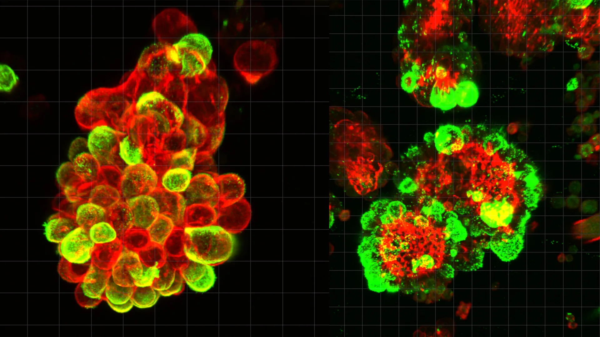
Ovarian cancer has long held a reputation as one of the most formidable challenges in oncology, frequently referred to as a "silent killer" due to its ability to proliferate extensively before clinical symptoms emerge. While most cancers spread through the bloodstream or lymphatic system, ovarian cancer utilizes a unique and aggressive method of movement within the abdominal cavity. A groundbreaking study led by researchers at Nagoya University in Japan has finally decoded the biological mechanism behind this rapid progression. Published in the journal Science Advances, the research reveals that ovarian cancer cells do not act in isolation; instead, they hijack healthy mesothelial cells—the protective lining of the internal organs—to act as "scouts" and "drills" that facilitate the invasion of new tissues. This symbiotic, yet deadly, partnership results in the formation of hybrid cell clusters that are significantly more resilient to conventional chemotherapy than independent cancer cells.
The Mystery of Transcoelomic Spread
For decades, oncologists have observed that ovarian cancer behaves differently from breast or lung cancers. In those diseases, malignant cells typically enter the circulatory system, allowing doctors to potentially detect them through blood-based biomarkers or "liquid biopsies." Ovarian cancer, however, primarily spreads through the transcoelomic route. Malignant cells detach from the primary tumor and enter the peritoneal fluid—a naturally occurring lubricating liquid that fills the space between abdominal organs.
Because this fluid is constantly in motion, driven by the physical act of breathing and digestive movements, cancer cells are carried like driftwood to various sites, including the liver, diaphragm, and intestinal lining. Until now, the scientific community lacked a clear understanding of how these floating cells successfully anchored themselves to new organs and why they were able to penetrate healthy tissue so efficiently. The Nagoya University study identifies the missing link: the active recruitment and transformation of mesothelial cells.
The Recruitment of Biological Accomplices
The research team, led by Dr. Kaname Uno, began their investigation by analyzing abdominal fluid, known as ascites, taken directly from patients diagnosed with ovarian cancer. Using high-resolution microscopy and single-cell genetic analysis, the team discovered that the cancer cells were rarely traveling alone. Instead, they were found in compact, spherical clusters consisting of both malignant cells and mesothelial cells.
Mesothelial cells are a vital part of the body’s defense; they form the mesothelium, a thin, slippery membrane that lines the thoracic and abdominal cavities, protecting organs from friction. However, the study found that ovarian cancer cells release a potent signaling molecule called Transforming Growth Factor-beta 1 (TGF-β1). When mesothelial cells, which have naturally shed from the abdominal lining, come into contact with this protein, they undergo a radical transformation.
Once "recruited" by the cancer cells, these mesothelial cells do not merely act as passengers. They become the primary drivers of tissue invasion. The study estimated that approximately 60% of all cancer cell clusters in the abdominal fluid contained these hijacked mesothelial cells. This partnership creates a hybrid entity that is far more dangerous than the sum of its parts.
Invadopodia: The Biological Drills of Ovarian Cancer
One of the most significant findings of the Nagoya University study is the identification of "invadopodia." These are specialized, spike-like protrusions that develop on the surface of the transformed mesothelial cells. While the cancer cells themselves remain relatively passive during the initial stages of attachment, the recruited mesothelial cells use these invadopodia to physically drill into the protective layers of distant organs.
Dr. Uno explained that the cancer cells essentially "outsource" the hard work of invasion. "The cancer cells undergo minimal genetic or molecular changes during this phase," Dr. Uno noted. "Instead, they manipulate the mesothelial cells to create openings in the tissue. The cancer cells then simply migrate through the pathways that their accomplices have cleared."
This mechanism explains why ovarian cancer can colonize the entire abdominal cavity with such speed. The hybrid spheres act like armored units: the mesothelial cells provide the "engine" and the "drill," while the cancer cells provide the "blueprint" for growth. Furthermore, the compact structure of these hybrid spheres provides a physical shield, making it harder for chemotherapy drugs to penetrate and kill the malignant cells at the center.
A Physician’s Motivation: The Story of Dr. Kaname Uno
The impetus for this research was deeply personal for the lead author. Before transitioning into full-time molecular research, Dr. Kaname Uno spent eight years as a practicing gynecologist. During his clinical tenure, he witnessed firsthand the devastating speed of ovarian cancer.
The study highlights one specific case that haunted Dr. Uno’s career: a patient who had undergone routine gynecological screenings and received a clean bill of health. Only three months later, she returned to the clinic with advanced, Stage IV ovarian cancer. Despite the best available treatments, the disease had already colonized her entire abdomen, and she passed away shortly thereafter.
"The existing diagnostic tools failed her," Dr. Uno stated. "There was no way to see the cancer coming because we didn’t understand the mechanism of its movement." This tragedy motivated him to join the Graduate School of Medicine at Nagoya University to investigate the "floating phase" of ovarian cancer cells—the window of time between detachment from the primary tumor and the colonization of new organs.
Chronology of the Research and Methodology
The study was conducted over several years, utilizing a multi-disciplinary approach to validate the findings. The chronology of the discovery followed a rigorous scientific path:
- Clinical Sampling: The team collected and preserved abdominal fluid from a diverse cohort of ovarian cancer patients at various stages of the disease.
- Advanced Microscopy: Using live-cell imaging, the researchers observed the physical interaction between cancer cells and mesothelial cells, documenting the formation of the hybrid spheres in real-time.
- Genomic Analysis: Through single-cell RNA sequencing, the team identified the specific gene expressions that were activated when TGF-β1 was released. They confirmed that the mesothelial cells were being reprogrammed at a molecular level to produce the proteins necessary for invadopodia.
- In Vivo Validation: The researchers utilized mouse models to test whether blocking the TGF-β1 signal would slow the spread of the disease. The results were clear: without the ability to recruit mesothelial cells, the cancer’s ability to invade distant organs was significantly hampered.
Supporting Data and Statistical Context
Ovarian cancer remains the deadliest gynecological malignancy worldwide. According to global health statistics, the five-year survival rate for ovarian cancer is approximately 49%, but this drops significantly to below 30% if the cancer is diagnosed at an advanced stage (Stage III or IV). Unfortunately, due to the lack of early symptoms and the rapid spread mechanism identified in this study, nearly 75% of patients are diagnosed at these later stages.
The Nagoya University data showing that 60% of cancer clusters are hybrids provides a new metric for pathologists. In the past, abdominal fluid analysis focused primarily on the presence of malignant cells. This research suggests that the presence and concentration of mesothelial-cancer hybrids could serve as a much more accurate predictor of a patient’s prognosis and the likely aggressiveness of the disease.
Implications for Future Treatment and Diagnostics
The discovery of the mesothelial "scout" mechanism opens several new doors for the treatment of ovarian cancer, which has seen relatively few breakthroughs in the last two decades.
1. Targeted Disruption of the TGF-β1 Pathway
Current chemotherapy protocols, such as the use of carboplatin and paclitaxel, focus on killing rapidly dividing cells. However, they do not address the signaling pathways that allow cancer cells to recruit healthy cells. Future therapies could involve inhibitors that block the TGF-β1 receptor, effectively "blinding" the cancer cells and preventing them from enlisting mesothelial accomplices.
2. Preventing Hybrid Sphere Formation
By understanding the "glue" that holds these spheres together, researchers can look for ways to break the clusters apart while they are still floating in the abdominal fluid. Once separated, cancer cells are more vulnerable to the body’s immune system and to standard chemotherapy.
3. Enhanced Monitoring and Liquid Biopsies
The study suggests that monitoring abdominal fluid for these hybrid clusters could become a standard part of follow-up care for patients in remission. If these clusters can be detected early through minimally invasive procedures, doctors may be able to intervene before new tumors take root on the liver or intestines.
Broader Impact on the Scientific Community
The findings from Nagoya University have resonated across the global oncological community. By shifting the focus from the cancer cell itself to the "microenvironment" and the interaction with healthy cells, the study aligns with a growing movement in cancer research that views tumors as complex ecosystems rather than isolated masses of rogue cells.
As Dr. Uno and his colleagues continue their work, the goal is to translate these laboratory findings into clinical trials. For the thousands of women diagnosed with ovarian cancer each year, this research offers a glimmer of hope that the "silent killer" may finally be stripped of its most effective weapon. The ability to predict and block the rapid abdominal spread of the disease could transform ovarian cancer from a terminal diagnosis into a manageable condition, potentially saving countless lives through earlier detection and more precise, targeted interventions.
