
 By George Ongkojoyo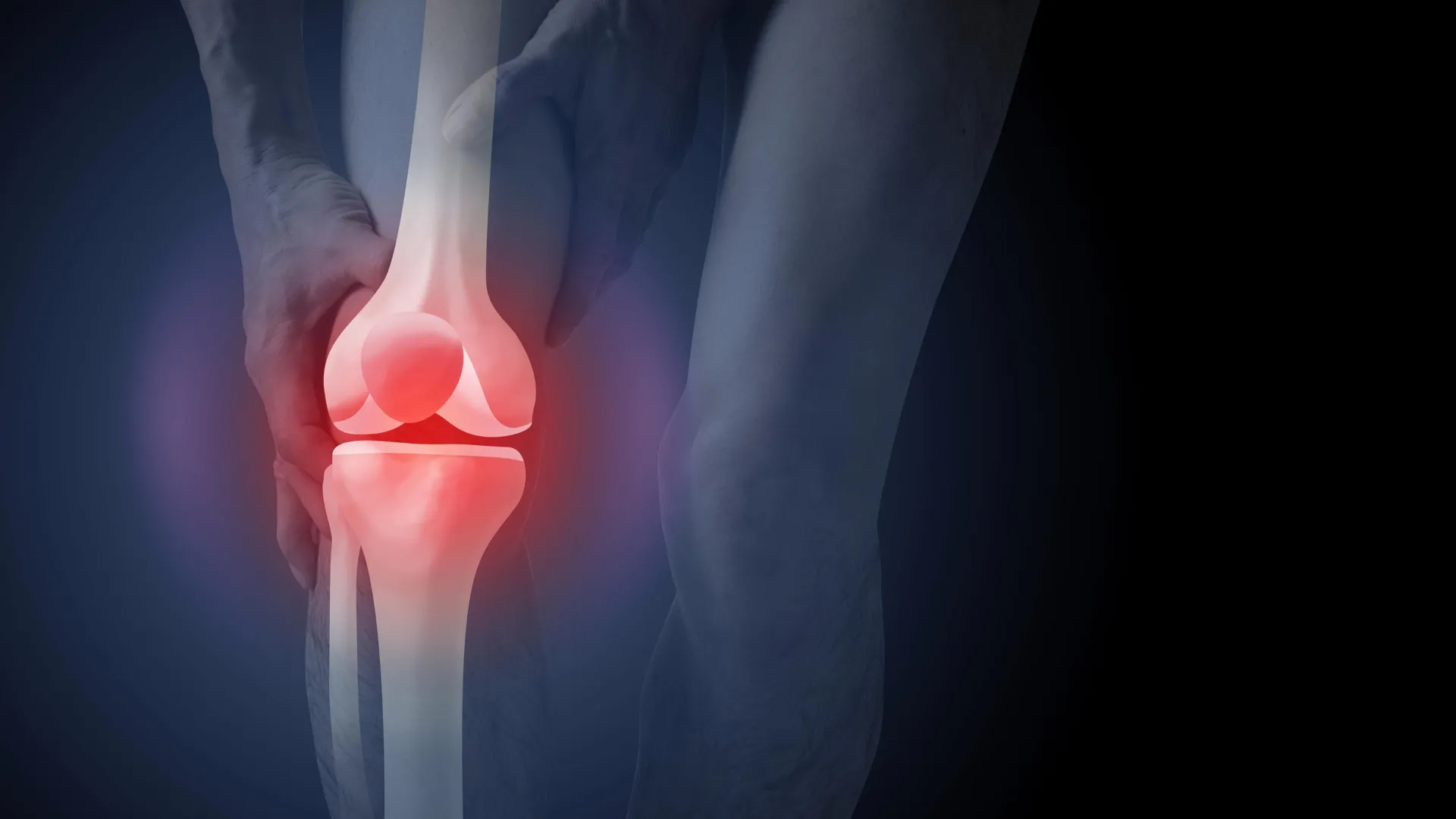
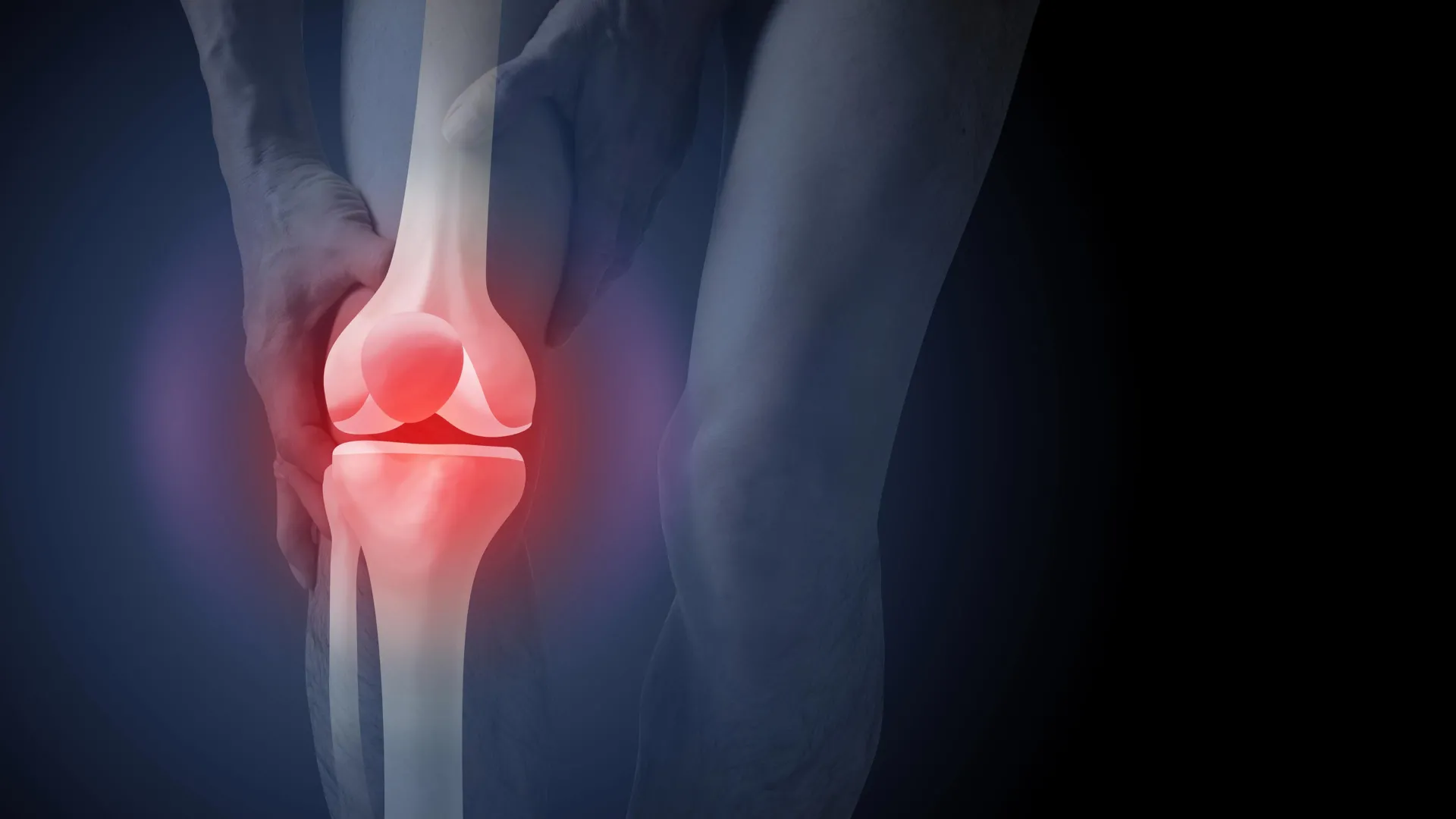
The clinical landscape for managing knee osteoarthritis in Australia is facing a significant challenge as new research highlights a disconnect between established medical guidelines and frontline diagnostic practices. Despite clear recommendations from peak health bodies stating that routine X-rays are not required for a diagnosis, nearly 50% of new patients presenting with knee pain to general practitioners (GPs) are still being referred for imaging. This trend is not merely a matter of administrative inefficiency; a groundbreaking study published in PLOS Medicine reveals that the use of X-rays profoundly alters patient perception, fueling a 36% increase in the perceived need for invasive surgery and heightening fears regarding physical activity.
As the Australian healthcare system grapples with the rising costs of chronic musculoskeletal conditions, the implications of this study suggest that a shift toward clinical-based diagnosis could save millions of dollars and improve patient outcomes. Currently, osteoarthritis imaging alone costs the national health system approximately A$104.7 million annually, while the broader hospital services related to the condition—largely driven by joint replacements—reached a staggering A$3.7 billion in the 2020–21 financial year.
The Disconnect Between Guidelines and Practice
For years, the Australian Commission on Safety and Quality in Health Care has maintained that knee osteoarthritis should be diagnosed through a "clinical diagnosis." This approach relies on a patient’s medical history, age, and specific symptoms rather than structural imaging. According to the Clinical Care Standard, a diagnosis can be confidently made if a patient is aged 45 or older and experiences activity-related joint pain with either no morning stiffness or stiffness that resolves within 30 minutes.
However, the reality in GP clinics across the country tells a different story. Data indicates that a significant portion of the medical community continues to rely on X-rays as a primary diagnostic tool. This reliance is often driven by a combination of factors, including patient expectations, the misconception that imaging is a prerequisite for high-quality care, and "defensive medicine," where doctors order tests to ensure no structural anomaly is missed.
The consequences of this over-reliance are now being quantified. The new study, which surveyed 617 participants across Australia, used a randomized controlled design to observe how different diagnostic methods influenced patient beliefs. Participants were assigned to watch videos of hypothetical consultations. One group received a diagnosis based on clinical symptoms alone, while two other groups were diagnosed using X-rays—one group was shown their "damaged" joint images, and the other was not.
The Psychological Impact of Seeing "Wear and Tear"
The findings of the study were stark. Participants who were shown X-ray images of their knees reported a 36% higher belief that they would eventually require knee replacement surgery compared to those diagnosed clinically. Furthermore, these individuals expressed a significantly higher "fear of movement." They were more likely to believe that exercise and daily physical activities would further damage their joints, leading to a "vicious cycle" of inactivity, weight gain, and worsening symptoms.
Researchers point to the "nocebo effect" of medical imaging in chronic pain management. When a patient sees an X-ray showing bone-on-bone contact or osteophytes (bone spurs), they often internalize a "mechanical" view of their body. The term "wear and tear" is frequently used by both patients and clinicians, yet it is increasingly viewed as a harmful misnomer. Research has long shown that the structural changes visible on an X-ray do not accurately correlate with a person’s level of pain or physical disability. Many individuals with significant joint changes experience very little pain, while others with "normal" X-rays report debilitating symptoms.
By focusing on the image rather than the person, the diagnostic process may inadvertently convince patients that their joints are "broken" and can only be fixed by a surgeon. This psychological shift often overshadows the most effective treatments for osteoarthritis: education, weight management, and structured exercise programs.
The Economic Burden of Osteoarthritis in Australia
The financial ramifications of over-imaging and the subsequent push toward surgery are substantial. In the 2021–22 period, more than 53,000 Australians underwent knee replacement surgery due to osteoarthritis. While these surgeries are life-changing for patients with end-stage disease who have exhausted all other options, experts worry that many are being performed prematurely.
The A$3.7 billion spent on osteoarthritis hospital services represents a significant portion of the national health budget. Reducing the number of unnecessary X-rays could act as a "gatekeeper" function, preventing the psychological priming that leads patients to demand surgical interventions before trying conservative management.
Furthermore, the A$104.7 million spent annually on imaging could be redirected toward subsidized physiotherapy, weight-loss programs, and community-led exercise initiatives like the GLAD (Good Life with Osteoarthritis in Denmark) program, which has seen high success rates in Australia.
A Timeline of Evolving Diagnostic Standards
The shift away from imaging is not a new concept, but it has gained momentum over the last decade as evidence-based medicine has matured.
- 2010–2015: International guidelines begin de-emphasizing X-rays for initial osteoarthritis diagnosis, noting the poor correlation between imaging and pain.
- 2017: The Australian Commission on Safety and Quality in Health Care releases the Osteoarthritis of the Knee Clinical Care Standard, explicitly stating that X-rays are not recommended for routine diagnosis.
- 2020: Data reveals that despite these standards, imaging rates remain high, and knee replacement surgeries continue to climb at rates that outpace population growth.
- 2024: The latest study in PLOS Medicine provides the "missing link" by demonstrating that X-rays are not just a neutral diagnostic tool but a psychological catalyst for surgical demand and fear-avoidance behavior.
Professional Reactions and the Path Forward
Health advocates and researchers are calling for a multi-pronged approach to change the current trajectory. The Royal Australian College of General Practitioners (RACGP) has previously noted that changing clinical habits requires more than just publishing guidelines; it requires a shift in how doctors communicate with patients.
"We need to move the conversation away from ‘what does the bone look like’ to ‘how does the joint function,’" suggests the research team behind the PLOS Medicine study. They argue that when a GP explains a clinical diagnosis, they have an opportunity to empower the patient. By focusing on muscle strength, joint loading, and lifestyle factors, the GP can frame osteoarthritis as a manageable condition rather than a degenerative death sentence for the knee.
From a patient perspective, the desire for an X-ray is understandable. In a culture that prizes high-tech medical intervention, a clinical diagnosis can sometimes feel like "lesser" care. The study found that participants were actually slightly more satisfied with an X-ray-based diagnosis than a clinical one, even if it led to worse psychological outcomes. This highlights a significant "expectation gap" that public health campaigns must address.
Broader Implications for Chronic Pain Management
The findings regarding knee osteoarthritis mirror similar trends in the treatment of chronic back and shoulder pain. In those fields, research has also shown that early imaging often leads to worse long-term outcomes, more invasive procedures, and higher levels of patient anxiety. By seeing "bulging discs" or "rotator cuff tears" on a screen, patients often become too afraid to engage in the very movements required to heal.
Reducing unnecessary medical radiation is another secondary benefit of adhering to clinical diagnosis standards. While a single knee X-ray involves a low dose of radiation, the cumulative effect of unnecessary imaging across a population of millions is a relevant public health consideration.
As Australia’s population ages and the prevalence of obesity—a major risk factor for osteoarthritis—continues to rise, the pressure on the healthcare system will only increase. Transitioning to a diagnosis model that prioritizes clinical symptoms over structural imaging offers a rare "win-win" scenario: it reduces the financial burden on the state while protecting patients from the anxiety and risks associated with unnecessary surgical pathways.
Summary of Recommendations for Patients
For Australians experiencing knee pain, the latest evidence suggests several key takeaways:
- Trust the Clinical Assessment: If a GP diagnoses osteoarthritis based on age and symptoms without an X-ray, they are following the highest standard of evidence-based care.
- Question the Need for Imaging: Patients are encouraged to ask their doctors how an X-ray will change their treatment plan. If the plan remains "exercise and weight management," the X-ray is likely unnecessary.
- Prioritize Conservative Management: Surgery should be viewed as a last resort. Programs focusing on strengthening the muscles around the knee and managing body weight are the first-line treatments and often yield results comparable to surgery without the risks of infection or blood clots.
- Understand the Joint: Osteoarthritis is not "wear and tear" but a process of the joint working to repair itself. Movement is "lube for the joint," and staying active is essential for long-term health.
The study concludes that by reforming how we diagnose this common condition, Australia can foster a more resilient patient population and a more sustainable healthcare system. The focus must return to the patient’s experience of pain and function, rather than the static, often misleading images of their bones.
