
 By Nana O
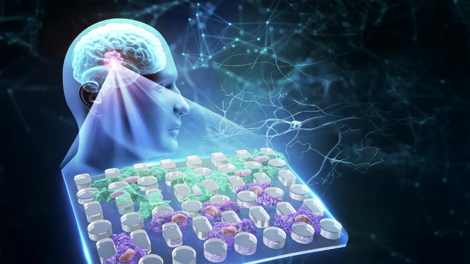
Glioblastoma, a relentless and devastating form of brain cancer, has long confounded medical science due to its aggressive nature and resistance to treatment. Now, groundbreaking research has unveiled a surprising and counterintuitive factor that may significantly contribute to its invasiveness: the varying viscosity of the fluid surrounding the tumor. Scientists have discovered that the fluid at the outer edges of glioblastoma tumors is considerably thicker than that found at their centers. This gradient of resistance, rather than simply impeding cancer cell movement, appears to be a potent catalyst for cellular adaptation, equipping these cells with enhanced abilities to infiltrate and destroy healthy brain tissue.
A Novel Approach to Understanding Cellular Mechanics
For decades, cancer research has predominantly focused on the intricate biochemical signaling pathways and the physical stiffness of the tumor microenvironment as key drivers of cancer progression and metastasis. The role of fluid viscosity, the measure of a fluid’s resistance to flow, has largely been overlooked. This oversight is particularly significant in aggressive brain tumors like glioblastoma, where the physical forces exerted on migrating cancer cells are substantial.
The disparity in fluid viscosity within a glioblastoma tumor is stark. Studies have indicated that the fluid at the tumor’s advancing edge can be up to eight times more viscous than the fluid within the necrotic, dead tissue at its core. As glioblastoma cells push outwards, they encounter an increasingly formidable barrier, a physical challenge that traditional research models have struggled to accurately replicate.
Conventional microfluidic devices, often employed to study cellular behavior under controlled conditions, have presented significant limitations in this context. Their enclosed channels can restrict the flow of oxygen and nutrients essential for long-term cell culture, a critical factor for observing sustained adaptation. Furthermore, friction against the channel walls introduces extraneous forces that can skew experimental results, making it difficult to isolate the specific impact of fluid viscosity. These limitations have hindered the ability of researchers to understand how prolonged exposure to viscous environments might fundamentally alter glioblastoma cell behavior.
Pioneering the Open Microfluidic Platform
To overcome these challenges, a team of researchers from Chongqing General Hospital and Chongqing University in China embarked on developing an innovative, open microfluidic platform. This novel device, detailed in a recent publication in Microsystems & Nanoengineering, was specifically designed to mimic the complex physical conditions encountered by glioblastoma cells at the tumor’s invasive front.
The platform’s ingenious design features a two-layer open microfluidic membrane equipped with a removable cap and an array of precisely engineered microscopic pillars. This configuration allows for meticulous control over when cancer cell migration commences and provides an unobstructed view of cellular dynamics, including the crucial deformation of cell nuclei as they navigate confined spaces.
A key advantage of this open system is its capacity for extended cell culture, allowing researchers to observe cellular responses for up to one month. This extended observation period is crucial for investigating the lasting effects of mechanical stress, particularly from fluid viscosity, on glioblastoma cell invasiveness. By isolating the influence of viscosity from other mechanical factors, the researchers aimed to determine if prolonged exposure to thicker fluids could induce persistent changes in these aggressive cancer cells.
Unveiling the Secrets of Viscous Adaptation
In their experiments, the scientists utilized two well-established human glioblastoma cell lines: U-251 and LN-229. These cells were cultured for a month in a specially formulated viscous fluid, designed to accurately represent the rheological properties of the fluid found at the tumor’s invasive edge, with a viscosity of approximately 7.1 centipoise (cP).
The results of these experiments were striking and, to some extent, paradoxical. When the adapted cells were subsequently introduced to the microfluidic chip, they exhibited significantly enhanced migratory capabilities. They not only traveled faster but also covered greater distances compared to untreated control cells. This finding directly challenged the conventional understanding that increased resistance would inherently slow down cellular movement. Instead, it suggested that glioblastoma cells were actively adapting to and even benefiting from this viscous environment.
The Transformation: Smaller, More Flexible, and Mechanically Literate Cells
Microscopic analysis provided crucial insights into the mechanisms behind this enhanced invasiveness. The cells that had undergone prolonged exposure to the viscous fluid had undergone remarkable physical transformations. They had become visibly smaller and demonstrably more flexible, enabling them to more effectively navigate and squeeze through the narrow interstitial spaces within the microfluidic device’s pillar array.
During this process of squeezing through constricted passages, the cell nuclei exhibited significant deformation, a visual cue indicating the immense mechanical forces at play. Furthermore, the researchers observed a notable accumulation of YAP, a critical protein known to be involved in sensing mechanical forces within cells, within the nuclei. The presence of YAP in the nucleus is a well-established indicator that cells have activated intracellular pathways responsible for responding to and adapting to physical stress. This suggests a sophisticated cellular response to the mechanical challenges posed by the viscous tumor microenvironment.
Interestingly, while both glioblastoma cell lines displayed similar physical adaptations in terms of shape and mobility, their underlying molecular responses revealed unexpected diversity. The U-251 cells underwent a form of epithelial-mesenchymal transition (EMT)-like reprogramming, a process known to be associated with enhanced cancer cell motility and invasiveness. This reprogramming was accompanied by the activation of key genes implicated in cancer invasion, including CD44, FN1, and MMP9.
In contrast, the LN-229 cells achieved increased flexibility and mobility without exhibiting substantial, lasting alterations in their gene expression profiles. This divergence highlights that glioblastoma cells may acquire comparable invasive characteristics through distinct biological pathways, underscoring the complexity of this formidable cancer.
Further investigation using Western blotting confirmed that certain protein alterations induced by the viscous environment persisted even after the cells were transferred back into a fluid of normal viscosity. This observation strongly suggests that some of the adaptations are not transient responses to immediate surroundings but rather represent a more enduring, "mechanical memory" within the cells.
A "Cellular Memory" of Mechanical Stress
Perhaps the most profound and unexpected finding of this research is the implication of a "cellular memory" in glioblastoma. The researchers initially hypothesized that fluid viscosity would primarily act as a physical impediment. However, their experiments demonstrated that prolonged exposure to viscous conditions could induce lasting changes in cellular behavior, even when the original mechanical stimulus was removed.
This phenomenon points towards a form of mechanical memory, where cells retain adaptations forged by earlier physical stresses. The open design of the microfluidic chip was instrumental in uncovering this effect. By effectively decoupling the resistance imparted by the fluid itself from the confinement imposed by channel walls, the researchers could isolate and study the pure effect of viscosity without the confounding mechanical interference often present in traditional microfluidic experiments.
The observation of altered nuclear shape and migration patterns after weeks of exposure provided compelling evidence that glioblastoma cells possess a remarkable capacity to adapt to their mechanical environment in persistent ways. The contrasting molecular responses observed between the two cell lines were particularly illuminating. Although both exhibited similar physical transformations, only one line showed widespread changes in gene activity. This suggests that glioblastoma cells may achieve enhanced invasiveness through a variety of biological mechanisms, all converging on a similar functional outcome.
Implications for Future Cancer Therapies and Research Tools
Beyond elucidating an overlooked factor in glioblastoma invasion, the researchers believe their pioneering microfluidic platform holds significant promise as a valuable tool for investigating novel cancer therapies. The open microfluidic platform’s compatibility with standard multi-well plates makes it readily integrable into existing laboratory workflows for cell culture, drug screening, and advanced imaging techniques. Its accessible design also facilitates direct cell staining and long-term observation without the clogging issues often encountered in closed microfluidic systems.
These characteristics position the platform as an ideal system for testing drugs designed to disrupt cellular pathways responsible for mechanosensing and mechanical force response. For glioblastoma, these findings suggest that highly viscous microenvironments within the tumor may actively select for and promote the growth of cancer cells that are pre-adapted for invasion. Consequently, therapeutic strategies targeting YAP signaling or the remodeling of the cytoskeleton—the intricate internal framework that governs cell shape and movement—could be particularly relevant when studied under conditions that more accurately reflect the in-vivo tumor environment.
The potential applications of this technology extend beyond glioblastoma. It could prove invaluable for studying other cancers where fluid viscosity varies significantly across different tumor regions. By examining how tumor cells adapt to these mechanical gradients, researchers could gain deeper insights into the mechanisms driving cancer invasiveness and identify patients whose tumors rely heavily on mechanical adaptation for their spread.
While further research is undoubtedly necessary to fully translate these laboratory findings into clinical applications and understand their direct relevance to glioblastoma patients, this discovery undeniably highlights a critical and previously underappreciated aspect of cancer biology. The physical environment surrounding tumor cells is not merely a passive backdrop but an active participant that can profoundly shape a cancer’s ability to invade and colonize healthy tissues. This paradigm shift in understanding the mechanical landscape of cancer opens new avenues for diagnostic and therapeutic intervention.
A Glimpse into Funding and Support
The groundbreaking research that illuminated the role of fluid viscosity in glioblastoma invasion was generously supported by several key institutions and funding bodies. The Chongqing Research Center for Glioma Precision Medicine and the Chongqing Key Laboratory of Neurodegenerative Diseases provided foundational institutional backing.
Significant financial contributions came from the National Natural Science Foundation of China, with grants awarded under numbers 82473430, 22304143, and 32501283. Additional support was provided by the Chongqing Talent Project (cstc2022ycjh-bgzxm0081) and the Natural Science Foundation of Chongqing (CSTB2025NSCQ-GPX1077, CSTB2025NSCQ-GPX0309).
Early-career researchers were supported through the Young Medical Talent Program of Chongqing (YXQN2025088) and the New Chongqing Project (CSTB2024YCJH-KYXM0115). Institutional research funding from Chongqing General Hospital (2025022102) and the Chongqing Special Funding for Postdoctoral Research Projects (2024CQBSHTB3131) also played a vital role. Furthermore, the Zhejiang Provincial Natural Science Foundation of China contributed with grant LQN25E050020, underscoring the collaborative and multi-faceted nature of this significant scientific endeavor.
