
 By Gabriel Paijo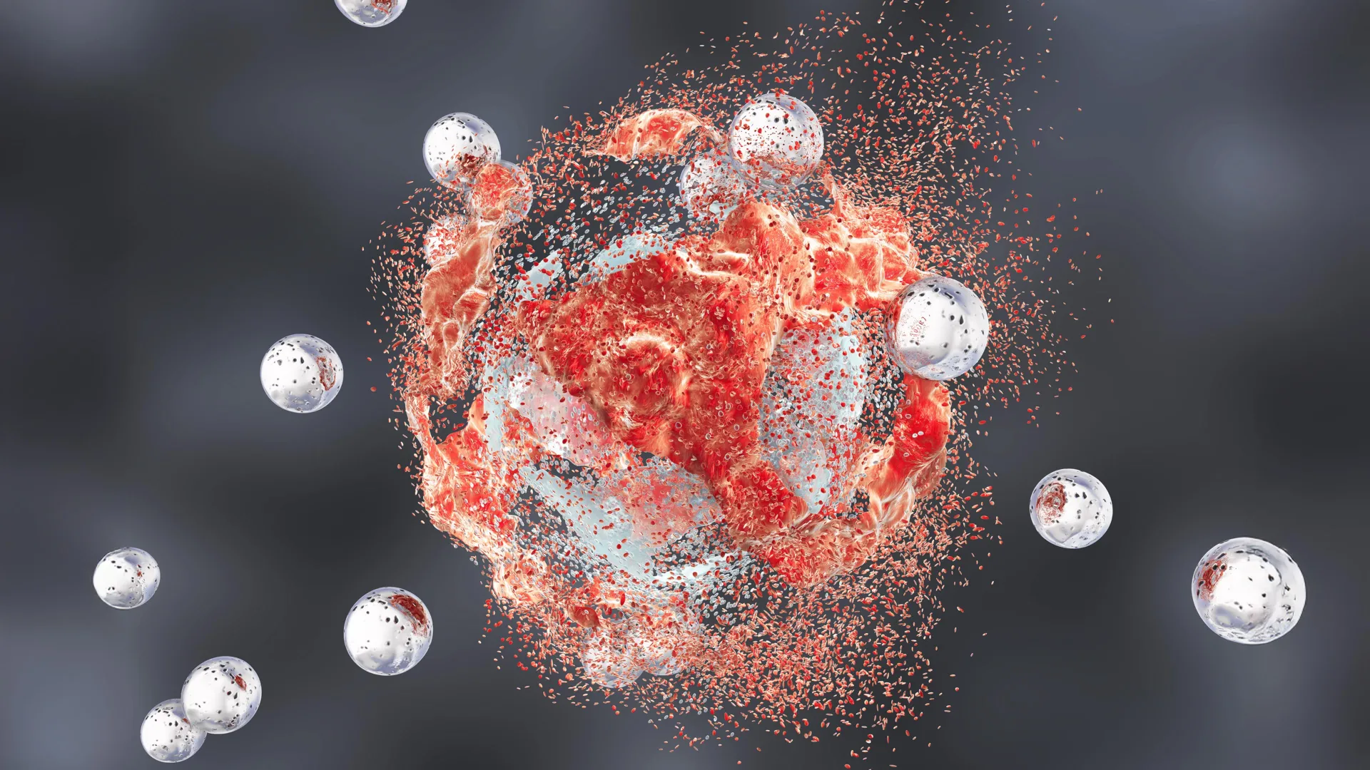
A multidisciplinary research team led by scientists at Weill Cornell Medicine and the Cornell Duffield College of Engineering has unveiled a sophisticated therapeutic platform utilizing engineered silica nanoparticles to treat aggressive prostate cancer. This preclinical study, published in the journal Cancer Research, details how these "ultrasmall" particles execute a two-pronged assault on malignancy: directly inducing a specialized form of cell death known as ferroptosis and simultaneously reprogramming the tumor’s immune environment to become more receptive to treatment. In experimental mouse models, the application of these particles, especially when combined with existing immunotherapies, resulted in significant tumor regression and complete remissions, marking a potential shift in how metastatic and treatment-resistant cancers are managed in the future.
The development of these particles, formally known as ultrasmall fluorescent core-shell silica nanoparticles or "Cornell Prime dots" (C’ dots), represents the culmination of over two decades of collaborative materials science and biomedical engineering. While C’ dots were originally conceived as highly precise imaging agents for surgical guidance, this latest research confirms that their biological utility extends far beyond visualization. By engineering the particles to target specific proteins on the surface of cancer cells, researchers have transformed a diagnostic tool into a potent therapeutic delivery system capable of dismantling the metabolic and immunological defenses of one of the most common and lethal cancers in men.
The Evolution of C’ Dots: From Imaging to Therapy
The journey of silica-based nanotechnology in medicine began with the search for safer, more stable contrast agents. Traditional dyes often suffer from rapid degradation or systemic toxicity. C’ dots, developed in the laboratory of Dr. Ulrich Wiesner at Cornell University, addressed these issues by encapsulating fluorescent molecules within a protective silica shell. Measuring less than 10 nanometers in diameter, these particles are small enough to circulate through the bloodstream and penetrate deep into tumor tissues, yet they are efficiently cleared by the kidneys, minimizing long-term accumulation in the body.
The transition from "seeing" tumors to "killing" them occurred as researchers noticed the peculiar ways these particles interacted with cellular machinery. Amorphous silica—a form of silicon dioxide found in various natural sources—is generally recognized as safe by the FDA for certain applications. However, at the nanoscale, these particles exhibit unique catalytic properties. The latest study demonstrates that when these particles are modified with specific targeting ligands, they do not merely sit within the tumor; they actively disrupt its internal stability.
Decoding the Mechanism: Ferroptosis and Oxidative Stress
One of the most significant findings of the study is the role of ferroptosis in the nanoparticle-driven destruction of prostate cancer cells. Unlike apoptosis, the most common form of programmed cell death which many cancer cells learn to evade, ferroptosis is an iron-dependent process characterized by the overwhelming accumulation of lipid peroxides. This process leads to the catastrophic oxidation of the fatty molecules that comprise the cell membrane, effectively causing the cell to rupture from the inside out.
The researchers discovered that C’ dots act as a "Trojan horse" for iron. Although the particles themselves are silica-based, their structure and surface chemistry allow them to sequester positively charged iron ions from the surrounding environment. Once the particles are internalized by prostate cancer cells via receptor-mediated endocytosis, the sudden influx of iron triggers a massive oxidative burst.
"The ability to induce ferroptosis is a game-changer," noted senior author Dr. Michelle Bradbury, the Endowed Professor of Imaging Research in Radiology at Weill Cornell Medicine. "Many aggressive cancers develop mutations that make them resistant to traditional chemotherapy-induced apoptosis. By triggering an entirely different pathway of cell death, we can bypass those resistance mechanisms and strike the tumor where it is most vulnerable."
Turning "Cold" Tumors "Hot": Immune System Reawakening
Beyond the direct killing of cancer cells, the silica nanoparticles addressed a major hurdle in prostate cancer treatment: the "cold" tumor microenvironment. Most prostate tumors are notoriously resistant to immunotherapy because they lack significant infiltration of active T cells. Instead, they are often populated by immunosuppressive cells that shield the cancer from the body’s natural defenses.
The study revealed that as the C’ dots induced ferroptosis, they also released signaling molecules that alerted the immune system. This process, often referred to as "immunogenic cell death," transformed the tumor environment from an immune-suppressed "cold" state into an immune-active "hot" state. The researchers observed a significant increase in the presence of activated T cells and cancer-fighting macrophages within the tumors following treatment.
Furthermore, the nanoparticles disrupted the metabolic pathways of the tumor. By altering how cancer cells process nutrients and energy, the C’ dots made the remaining tumor cells more susceptible to external attacks. This metabolic reprogramming is crucial because it prevents the tumor from adapting to the stress of the treatment, leading to more durable outcomes.
Experimental Data and Survival Metrics
To validate the efficacy of the C’ dots, the research team conducted a series of survival studies using mouse models of aggressive, metastatic prostate cancer. To ensure the particles reached their destination, they were functionalized with a targeting molecule that binds to Prostate-Specific Membrane Antigen (PSMA), a protein highly expressed on the surface of most prostate cancer cells.
The results of the combination therapy trials were particularly striking:
- Monotherapy: Mice treated with only the C’ dots or only standard immune checkpoint blockade (ICB) therapy showed modest improvements in survival, but the tumors eventually continued to grow.
- Dual Combination: When C’ dots were combined with ICB therapy, the researchers observed a synergistic effect. Four out of ten mice achieved complete remission, surviving indefinitely without evidence of disease.
- Triple Combination: The team added a third agent—a CSF-1R blockade designed to deplete immunosuppressive macrophages. This triple-therapy approach increased the complete remission rate to 50%, with five out of ten mice showing total tumor clearance.
The data suggests that the nanoparticles do not just kill cells; they create a "primed" environment where other drugs can perform more effectively. This synergy is vital for treating prostate cancer, where immunotherapy drugs like pembrolizumab have historically shown limited success when used as standalone treatments.
Safety Profiles and Biocompatibility
A primary concern with any nanotechnology is systemic toxicity. However, the C’ dots demonstrated a remarkable safety profile. Because the particles are engineered to be "ultrasmall," they follow a distinct pathway through the body. Unlike larger nanoparticles that tend to get trapped in the liver and spleen, C’ dots are small enough to be filtered by the kidneys and excreted in urine.
During the study, the researchers monitored the mice for signs of organ damage or systemic inflammation. While some particles were briefly detected in the spleen—a common occurrence for foreign materials in the bloodstream—there was no evidence of long-term toxicity. The targeting mechanism ensured that the highest concentrations of silica were found within the prostate tumors, sparing healthy tissues from the oxidative stress associated with ferroptosis.
Dr. Ulrich Wiesner, a co-corresponding author and professor of Materials Science and Engineering at Cornell, expressed a sense of wonder at the particles’ biocompatibility. "Silica is a fundamental part of our environment, found in the very foods we eat. It appears that our biology has an inherent way of interacting with these materials. We are only now beginning to understand how to harness that connection for therapeutic purposes."
Historical Context and Research Timeline
The success of this study is the result of a long-term collaboration that spans nearly two decades. The partnership between Dr. Bradbury’s clinical imaging expertise and Dr. Wiesner’s materials science background has led to multiple iterations of silica technology.
- Early 2000s: Initial development of C’ dots for fluorescent imaging in laboratory settings.
- 2011: The first human clinical trial for C’ dots as an imaging agent for melanoma, marking the first time the FDA approved such a nanoparticle for human use.
- 2018-2020: Discovery of the inherent therapeutic properties of silica, specifically its ability to induce nutrient deprivation and oxidative stress in certain cancer types.
- 2021-2023: Refinement of the PSMA-targeting platform and the initiation of the prostate cancer-specific studies funded by the Department of Defense and the National Cancer Institute.
- June 2024: Publication of the definitive study in Cancer Research outlining the dual-action mechanism and its potential for immunotherapy synergy.
Broader Implications for Oncology
The implications of this research extend beyond prostate cancer. The ability to induce ferroptosis and remodel the immune microenvironment could theoretically be applied to other "cold" tumors, such as pancreatic cancer or certain types of breast cancer. By changing the surface targeting molecule, researchers could redirect these silica "smart bombs" to various types of malignancies.
Dr. Jedd Wolchok, a study co-author and director of the Meyer Cancer Center at Weill Cornell Medicine, highlighted the clinical significance of the work. "In the field of oncology, we are constantly looking for ways to make immunotherapy work for more patients. This technology addresses the fundamental problem of immune exclusion. By using a nanoparticle to physically and chemically alter the tumor, we are opening the door for the immune system to do what it does best."
The study also underscores the importance of "metabolic oncology"—the burgeoning field that focuses on disrupting the specific energy needs of cancer cells. As C’ dots interfere with the iron metabolism and lipid stability of the tumor, they represent a new class of "metabolic therapeutics" that differ from traditional cytotoxic drugs.
Future Directions: Toward Human Clinical Trials
With the preclinical data showing such strong evidence of efficacy and safety, the research team is now preparing for the next phase of development. The transition from mouse models to human patients requires rigorous standardization of manufacturing and additional safety testing.
The team plans to evaluate the C’ dots in a Phase I clinical trial, focusing on patients with advanced prostate cancer who have exhausted standard treatment options. These trials will aim to determine the optimal dosing schedule and confirm whether the "cold-to-hot" immune transformation observed in mice translates to the human immune system.
Furthermore, the researchers are investigating whether the particles can be loaded with additional therapeutic payloads, such as radiopharmaceuticals or small-molecule drugs, to create an even more potent "all-in-one" treatment delivery system.
The study was supported by significant federal and private funding, including the Department of Defense and the Parker Institute for Cancer Immunotherapy. As the researchers move forward, the goal remains clear: to turn a tiny piece of silica into a life-saving intervention for the millions of men affected by prostate cancer worldwide. Through the convergence of engineering, radiology, and immunology, the C’ dot platform may soon represent a new paradigm in the ongoing effort to outmaneuver cancer’s complex defense systems.
