
 By Suherman Candra Maholtra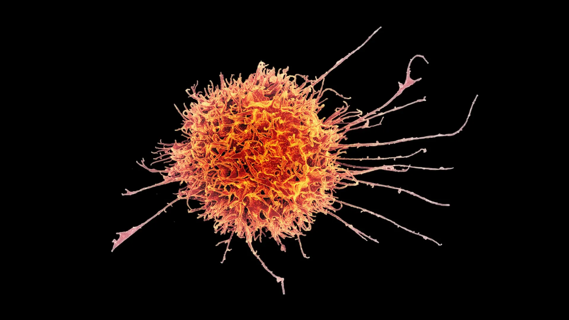
The landscape of oncology has been fundamentally altered over the last decade by the advent of cell-based immunotherapies, yet a significant barrier has remained: the stubborn resistance of solid tumors to these advanced treatments. While Chimeric Antigen Receptor (CAR) T-cell therapies have achieved remarkable success in treating hematologic malignancies, such as certain leukemias and lymphomas, they have largely faltered when faced with the dense, immunosuppressive environments of solid tumors. Researchers at Stanford Medicine, in collaboration with several leading institutions, have recently unveiled a breakthrough strategy that could bridge this gap. By reprogramming natural killer (NK) cells into a specialized "tissue-resident" state, the team has demonstrated a way to enable these immune cells to infiltrate, persist within, and ultimately destroy solid tumor masses.
The study, published in the journal Science Translational Medicine, outlines a methodology for transforming circulating NK cells—typically found in the blood—into a form that mimics the immune cells naturally found within organs like the lungs, liver, and skin. These engineered cells, known as cytotoxic tissue-resident NK cells, showed an unprecedented ability to navigate the hostile microenvironment of solid tumors in preclinical models. Led by senior author John Sunwoo, MD, the Edward C. and Amy H. Sewall Professor in the School of Medicine, the research suggests a future where cell therapy is not only more effective against common cancers but also more accessible as an "off-the-shelf" medical product.
The Challenge of the Solid Tumor Microenvironment
To understand the significance of the Stanford discovery, one must first look at why solid tumors—which account for approximately 90% of adult cancer cases—are so difficult to treat with current immunotherapies. Unlike blood cancers, where malignant cells circulate freely and are easily accessible to immune cells, solid tumors are physical fortresses. They are surrounded by a dense extracellular matrix and a network of stromal cells that act as a physical barrier.
Furthermore, solid tumors create an immunosuppressive microenvironment. They secrete signaling molecules that "exhaust" or deactivate T-cells and NK cells that manage to enter the tumor. One of the most potent of these signals is Transforming Growth Factor-beta (TGF-β). While TGF-β is essential for normal cell growth and wound healing, tumors hijack this protein to suppress the immune system. Previous attempts to use NK cells against solid tumors often failed because the cells would either remain in the peripheral blood or become dysfunctional once they encountered the high levels of TGF-β within the tumor tissue.
Engineering the "Goldilocks" Response
The Stanford team focused on a specific subset of immune cells: natural killer cells. Identified in the 1970s, NK cells are the "first responders" of the innate immune system. Unlike T-cells, which require a specific "key" to recognize a cancer cell (a process called antigen presentation), NK cells can recognize and kill abnormal cells instinctively. However, most NK cells used in therapy are derived from blood and lack the "homing" receptors needed to settle into solid tissues.
The researchers discovered that the key to creating effective tumor-fighting cells lay in the precise manipulation of TGF-β. Through a series of experiments isolating NK cells from human donors, the team found that the timing and intensity of TGF-β exposure acted as a biological switch.
"It’s a Goldilocks kind of thing," explained Dr. Sunwoo. If NK cells are exposed to too much TGF-β for too long, they become suppressed and lose their killing capacity. If they receive no signal, they remain blood-type cells that cannot enter tumors. The researchers found that a brief, intense burst of TGF-β—delivered via direct physical contact with short-lived tumor cells—triggered the cells to adopt a "tissue-resident" profile without losing their aggression.
These optimized cells expressed specific surface proteins, namely CD49a, CD103, and CD39. While CD49a and CD103 are markers of tissue residency, the presence of CD39 was identified as a hallmark of the most potent killers. These engineered cells were found to be packed with perforin and granzyme A, the molecular "ammunition" used to punch holes in cancer cell membranes and trigger programmed cell death.
Chronology of the Research and Preclinical Success
The development of this strategy followed a rigorous multi-year timeline, moving from basic cellular mapping to advanced mouse models. After identifying the ideal signaling "recipe" in the lab, the researchers moved to testing the cells in vivo.
In experiments involving mice with human-derived melanoma and head and neck squamous cell carcinoma, the results were definitive. The engineered tissue-resident NK cells didn’t just stay in the bloodstream; they migrated directly into the heart of the tumors. When compared to conventional NK cell treatments, the modified cells slowed tumor growth significantly more effectively.
The research reached its peak efficacy when the modified NK cells were paired with cetuximab, a monoclonal antibody currently used to treat certain head and neck and colorectal cancers. Cetuximab works by binding to the Epidermal Growth Factor Receptor (EGFR) on cancer cells, effectively "flagging" them for the immune system. In the study, mice receiving the combination therapy showed dramatic tumor suppression. By day 30 of the trial, while control groups showed advanced disease progression, the mice treated with the combination remained healthy, demonstrating a synergistic effect that could translate to higher survival rates in human patients.
Moving Toward an "Off-the-Shelf" Solution
One of the most significant implications of this research is the potential to revolutionize the logistics of cancer treatment. Currently, CAR T-cell therapy is an "autologous" process: a patient’s own cells must be harvested, genetically modified in a lab over several weeks, and then re-infused. This process is expensive, often costing hundreds of thousands of dollars, and the delay can be fatal for patients with rapidly progressing disease.
NK cells offer a different path. Unlike T-cells, they do not typically cause Graft-versus-Host Disease (GvHD), a dangerous condition where donor cells attack the recipient’s healthy tissues. This means NK cells can be harvested from healthy donors, modified, and used for any patient.
The Stanford team has already developed a scalable manufacturing process. According to the study, a single donor’s blood could provide enough material to produce approximately 20 doses of cytotoxic tissue-resident NK cells within a two-week window. These cells can be cryopreserved (frozen), allowing hospitals to keep them in stock.
"It would be almost an off-the-shelf drug," Dr. Sunwoo stated. This scalability could drastically reduce costs and eliminate the weeks-long waiting period currently associated with cell therapies, making life-saving treatments accessible to a much broader demographic of patients.
Broader Implications and Future Clinical Trials
The success of the Stanford study has sent ripples through the oncology community. By identifying the specific molecular markers (CD39) and the "Goldilocks" signaling threshold for TGF-β, the researchers have provided a blueprint for others in the field of synthetic biology.
The study also sheds light on the paradoxical nature of tissue-resident immune cells. In some contexts, like pregnancy, these cells are naturally immunosuppressive to protect the fetus. In the context of cancer, they can be either "friend or foe" depending on the signals they receive from the tumor. The ability to artificially steer these cells toward a "killer" phenotype represents a significant step forward in precision medicine.
Looking ahead, the Stanford team is moving rapidly toward human applications. They have applied for patents on the method for expanding these cytotoxic tissue-resident NK cells and are preparing for a Phase I clinical trial. This trial, which could begin as early as late 2024 pending FDA approval, will focus on patients with advanced squamous cell carcinoma of the head and neck—a group that currently has limited options when standard therapies fail.
The research was supported by a diverse coalition of funders, including the National Institutes of Health (NIH), the Tai Tsun Wu Research Fund for Natural Killer Cell Immunotherapy, and the Stanford Bio-X Fellowship. This collaborative effort, involving experts from Ohio State University and Washington University School of Medicine, underscores the multidisciplinary nature of modern cancer research.
While Dr. Sunwoo and his colleagues caution that mouse model successes do not always translate perfectly to human outcomes, the "proof of concept" established by this study provides a robust foundation. If the upcoming clinical trials mirror the preclinical results, the medical community may finally have the tools necessary to dismantle the physical and chemical defenses of solid tumors, turning the tide in the fight against the world’s most common and deadly cancers.
