
 By George Ongkojoyo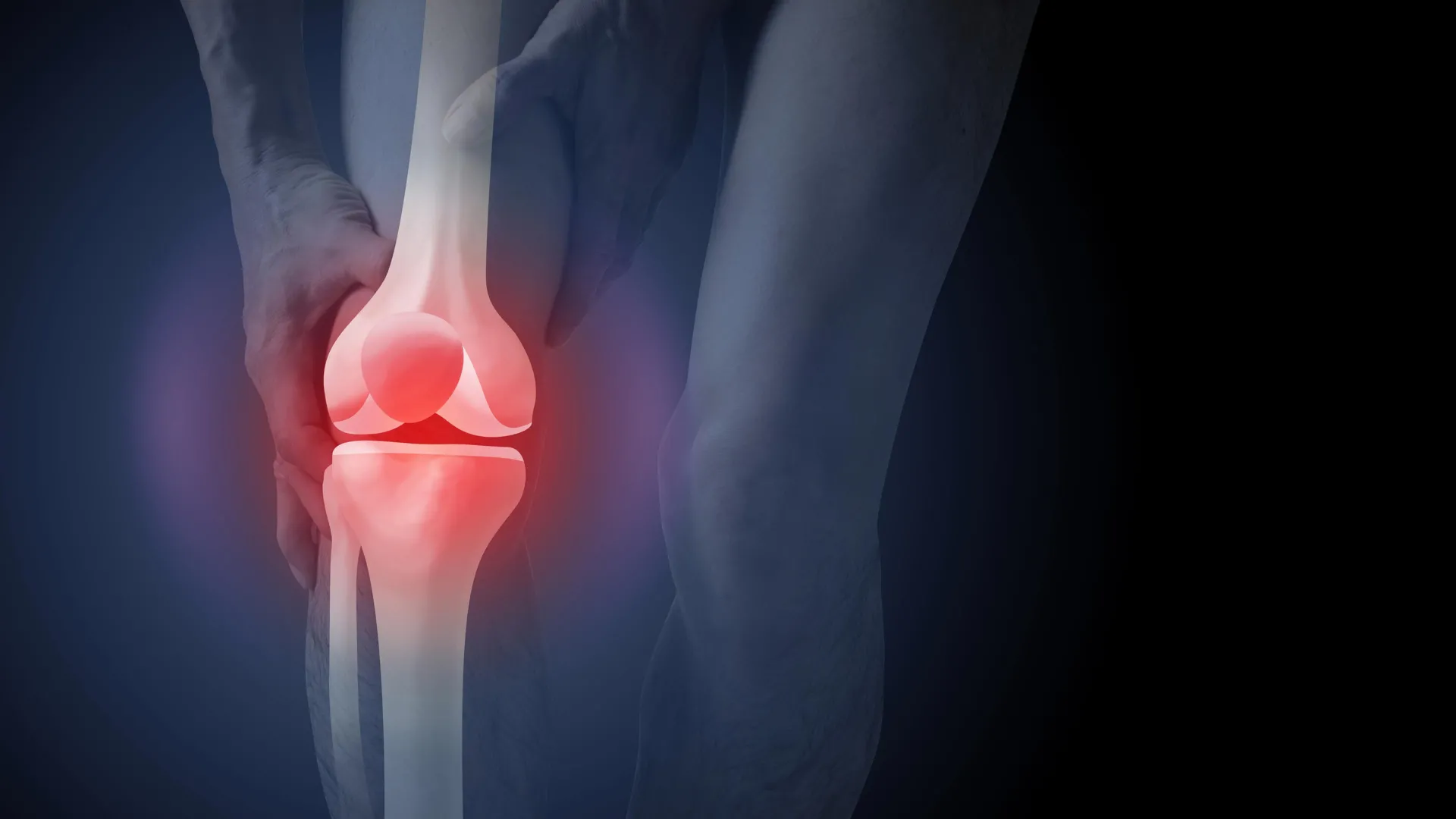
The diagnostic pathway for knee osteoarthritis in Australia is facing significant scrutiny following new research highlighting a disconnect between clinical guidelines and practitioner behavior. While the Australian Commission on Safety and Quality in Health Care explicitly advises against the use of routine imaging for the diagnosis of knee osteoarthritis, recent data indicates that nearly 50% of new patients presenting to general practitioners (GPs) with knee pain are still referred for X-rays or other imaging services. This trend is not merely a matter of administrative preference; it carries substantial economic weight and profound psychological implications for patients. Currently, the annual cost of imaging for osteoarthritis exceeds A$104.7 million, contributing to a broader financial burden on the national healthcare system that reached approximately $3.7 billion in the 2020–21 period.
A landmark study recently published in PLOS Medicine has provided empirical evidence that the use of X-rays in the diagnostic process fundamentally alters how patients perceive their condition. The findings suggest that seeing structural changes on an image can trigger a "nocebo" effect, where the patient’s belief in their physical vulnerability increases, leading them to pursue more invasive and potentially unnecessary interventions, such as total knee replacement surgery.
The Disconnect Between Imaging and Clinical Symptoms
For decades, the medical community has operated under the assumption that visualizing a joint is the most accurate way to assess its health. However, contemporary rheumatology and orthopedic science have debunked the notion that X-ray results correlate directly with patient experience. Research consistently demonstrates that the degree of "joint space narrowing" or the presence of "osteophytes" (bone spurs) seen on an X-ray does not accurately reflect the level of pain, disability, or future progression of the disease.
In many cases, an individual with significant structural changes visible on an X-ray may experience only minor discomfort, while another individual with a "clean" scan may suffer from debilitating chronic pain. This paradox is why major health bodies, including the National Institute for Health and Care Excellence (NICE) and the Royal Australian College of General Practitioners (RACGP), advocate for a "clinical diagnosis." This approach relies on a patient’s age—typically 45 years or older—and specific symptoms, such as activity-related joint pain and morning stiffness that resolves within 30 minutes.
Despite these clear directives, the habit of ordering imaging remains deeply ingrained in the Australian medical landscape. This is driven partly by a "better safe than sorry" mentality among clinicians and partly by patient expectations. Many patients believe that a diagnosis is not "official" or "valid" until it is confirmed by a picture of the bone, a misconception that the PLOS Medicine study sought to investigate and quantify.
Methodology and Findings of the PLOS Medicine Study
To understand the psychological impact of imaging, researchers conducted a controlled study involving 617 participants across Australia. The participants were randomly assigned to one of three experimental groups, each watching a video of a hypothetical consultation between a GP and a patient complaining of persistent knee pain.
In the first group, the doctor provided a clinical diagnosis based solely on the patient’s history and physical symptoms, explaining that X-rays were unnecessary. In the second and third groups, X-rays were utilized as the primary diagnostic tool. The key variable between these latter groups was whether the patient was actually shown the X-ray images during the consultation. Following the videos, participants were surveyed on their beliefs regarding treatment, the necessity of surgery, and their fear of physical activity.
The results were stark. Participants who were diagnosed via X-ray and shown their images reported a 36% higher perceived need for knee replacement surgery compared to the clinical diagnosis group. Furthermore, these individuals expressed significantly higher levels of "kinesiophobia"—the fear of movement—believing that exercise and physical activity would cause further "wear and tear" and permanent damage to their joints. This group was also more likely to express anxiety about their condition worsening over time.
Ironically, the study found that patients were slightly more satisfied with the X-ray-based diagnosis. This highlights a critical challenge for healthcare providers: patients often feel more "heard" or "thoroughly examined" when technology is involved, even when that technology leads to worse long-term psychological outcomes and riskier treatment choices.
The Economic Impact and the Rise of Knee Replacements
The financial implications of over-imaging are vast. Beyond the $104.7 million spent annually on the scans themselves, the X-ray often acts as the "gateway" to a high-cost surgical pathway. In 2021–22, more than 53,000 Australians underwent knee replacement surgery due to osteoarthritis. While these surgeries can be life-changing for patients with end-stage disease who have exhausted all other options, there is growing concern that many are being performed prematurely.
The total cost for hospital services related to osteoarthritis, driven largely by joint replacements, was estimated at $3.7 billion in 2020–21. This figure includes the costs of surgery, hospital stays, and immediate post-operative care. When considering the risks associated with major surgery—including blood clots, pulmonary embolisms, post-operative infections, and the reality that not all patients experience total pain relief—the push for non-surgical management becomes a matter of public health urgency.
The Biological Reality of Osteoarthritis
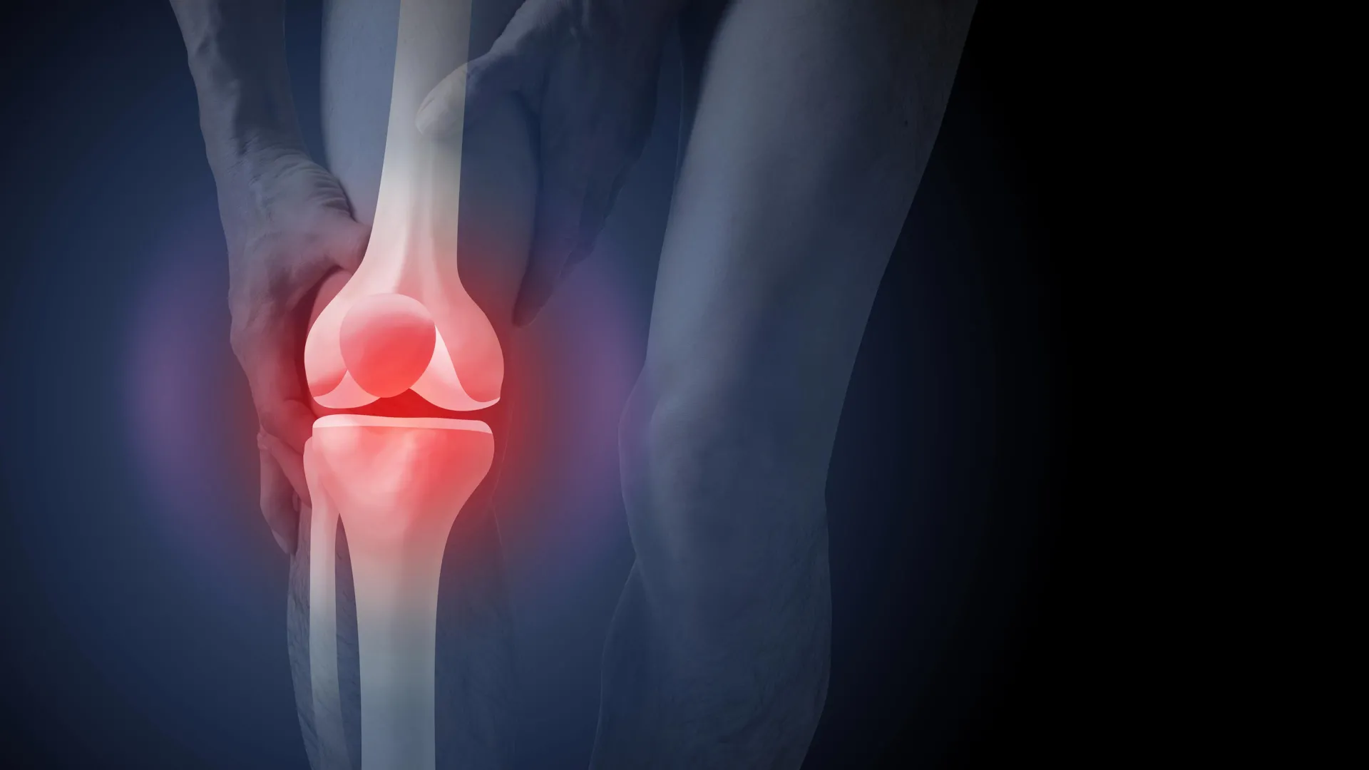
A central part of the problem is the pervasive "wear and tear" myth. The term implies that the knee is like a car tire that eventually loses its tread and must be replaced. In reality, osteoarthritis is a dynamic biological process. It is a condition of the entire joint—including the ligaments, muscles, bone, and synovial fluid—where the joint is working "extra hard" to repair itself in response to various stressors.
Factors that contribute to the development of osteoarthritis include a history of joint injury, high body weight, and genetic predispositions. However, the joint is a living tissue capable of adaptation. Modern treatment protocols emphasize that exercise does not "wear out" the joint; rather, it strengthens the supporting musculature, improves joint lubrication, and can actually reduce inflammation. By labeling the condition as "wear and tear" and showing patients images of bone spurs, clinicians inadvertently convince patients that their bodies are broken beyond the point of natural repair.
Rethinking the Patient Journey: From Scans to Strength
To address the findings of the study, health advocates are calling for a shift in how GPs communicate with patients. The goal is to move away from "low-value care"—treatments or tests that provide little benefit and may cause harm—toward evidence-based management.
Effective management of knee osteoarthritis typically involves a three-pronged approach:
- Education: Helping patients understand that their pain is not necessarily a sign of structural damage and that their joints are resilient.
- Exercise: Targeted physical therapy and strengthening exercises, such as those found in the GLA:D (Good Life with Osteoarthritis: Denmark) program, which has seen significant success in Australia.
- Weight Management: Reducing the mechanical load on the joint through sustainable lifestyle changes.
When these non-surgical options are prioritized, many patients find that their pain becomes manageable, their mobility improves, and the need for surgery is either delayed for decades or avoided entirely.
Chronology of Diagnostic Evolution
The shift away from imaging has been a slow process in the medical community. In the early 2000s, X-rays were the gold standard for any joint pain. By the 2010s, as longitudinal studies began to show the poor correlation between X-ray severity and pain levels, international guidelines began to change.
In 2018, the Australian Commission on Safety and Quality in Health Care released the Osteoarthritis of the Knee Clinical Care Standard, which explicitly stated that imaging should not be used for routine diagnosis. Despite this, the 2021–22 data shows that the "imaging-first" culture persists. The recent PLOS Medicine study (2024) serves as a critical update to this timeline, providing the psychological "missing link" that explains why over-imaging leads to over-surgery.
Broader Implications for the Healthcare System
The implications of this study extend beyond the knee. Similar trends have been observed in the treatment of lower back pain and shoulder impingements, where "incidental findings" on MRIs and X-rays—such as disc bulges or minor tears that are common in asymptomatic aging populations—lead to unnecessary anxiety and invasive procedures.
By reducing the number of unnecessary X-rays for knee pain, the Australian healthcare system could potentially save millions of dollars that could be redirected toward subsidized physiotherapy and exercise programs. More importantly, it would protect patients from the "fear-avoidance" cycle that often leads to sedentary lifestyles, weight gain, and a decline in overall cardiovascular health.
Medical professionals are now being urged to reconsider their diagnostic scripts. Instead of saying, "Let’s get an X-ray to see what’s going on," the recommendation is to say, "Based on your symptoms and age, we know this is osteoarthritis. The good news is that we don’t need an X-ray to start a treatment plan that will get you moving again."
As the Australian population ages and the prevalence of osteoarthritis rises, the sustainability of the healthcare system will depend on its ability to move away from expensive, image-driven models of care toward patient-centered, clinical management. The findings of the PLOS Medicine study provide a clear roadmap: to improve patient outcomes, we must first change what the patient sees and believes about their own body.
